
Medical image detection method based on deep learning and related equipment

A medical image and deep learning technology, applied in the fields of medical image detection, computer-readable media and electronic equipment, can solve the problems of low reliability of the lesion model and loss of three-dimensional volume data information, and achieve the effect of improving reliability

Active Publication Date: 2019-08-09
TENCENT TECH (SHENZHEN) CO LTD
View PDF4 Cites 43 Cited by
  • Summary
  • Abstract
  • Description
  • Claims
  • Application Information

AI Technical Summary

Problems solved by technology

This method is more suitable for image data such as mammary glands, but for the processing of CT (Computed Tomography, computerized tomography) images, it may lead to the loss of information of the three-dimensional volume data of CT images, making the established lesion model less reliable

Method used


Image

  • Medical image detection method based on deep learning and related equipment
  • Medical image detection method based on deep learning and related equipment
  • Medical image detection method based on deep learning and related equipment

Examples


Embodiment Construction

[0033] Example embodiments will now be described more fully with reference to the accompanying drawings. Example embodiments may, however, be embodied in many forms and should not be construed as limited to the examples set forth herein; rather, these embodiments are provided so that this disclosure will be thorough and complete and will fully convey the concept of example embodiments to those skilled in the art.

[0034] Furthermore, the described features, structures, or characteristics may be combined in any suitable manner in one or more embodiments. In the following description, numerous specific details are provided in order to give a thorough understanding of embodiments of the present disclosure. However, those skilled in the art will appreciate that the technical solutions of the present disclosure may be practiced without one or more of the specific details, or other methods, components, means, steps, etc. may be employed. In other instances, well-known methods, ap...


PUM


Abstract

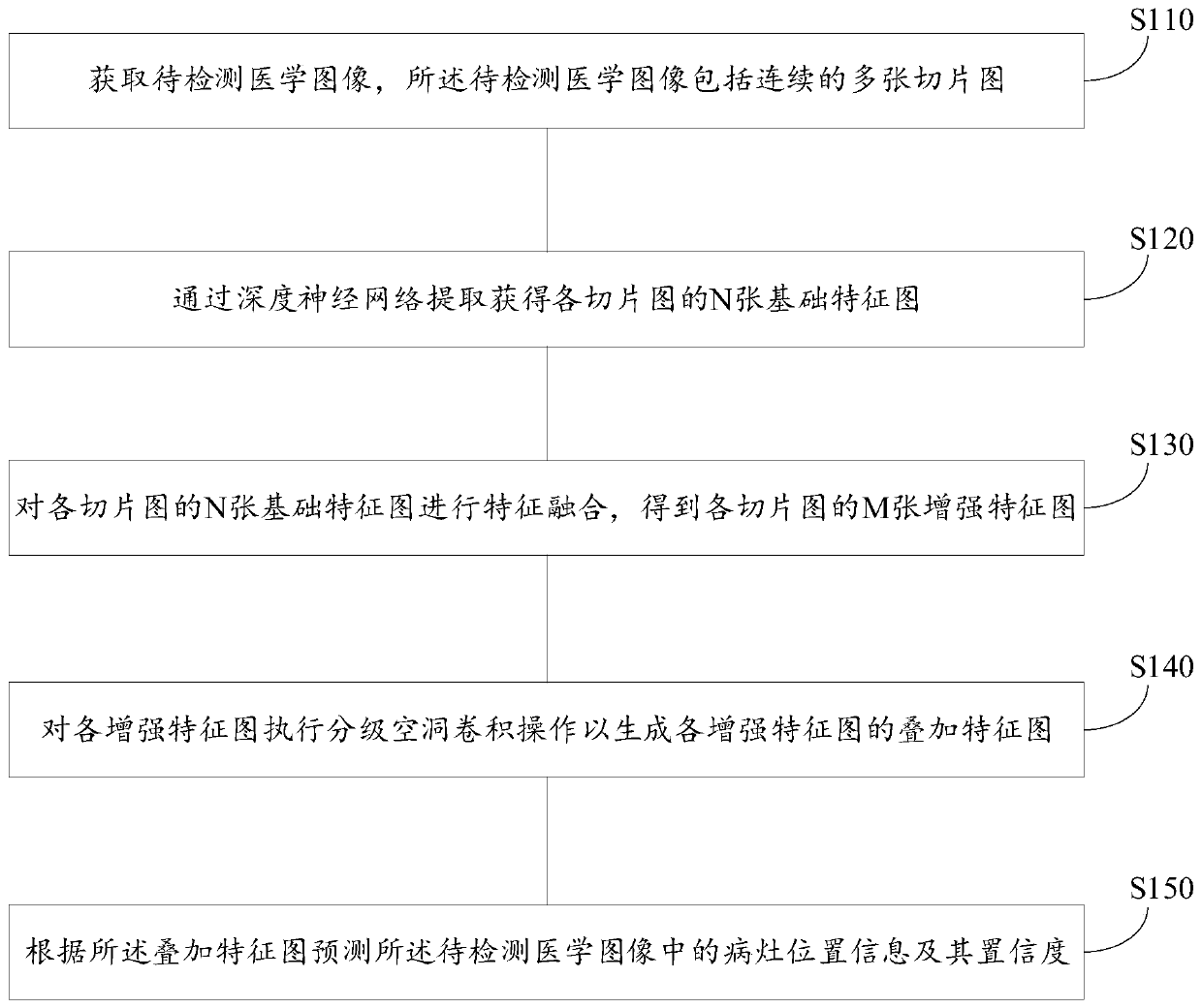
The embodiment of the invention provides a medical image detection method and device based on deep learning, a computer readable medium and an electronic device. The method comprises the steps of obtaining a to-be-detected medical image, wherein the to-be-detected medical image comprises a plurality of adjacent slice images; extracting to obtain N basic feature maps of each slice map through a deep neural network; performing feature fusion on the N basic feature maps of each slice map to obtain M enhanced feature maps of each slice map; performing a hierarchical void convolution operation on each enhanced feature map to generate a superposition feature map of each enhanced feature map; predicting focus position information and confidence coefficient thereof in the to-be-detected medical image according to the superposition feature map; wherein N and M are both positive integers greater than 1. According to the technical scheme provided by the embodiment of the invention, the target detection accuracy of the medical image can be improved.

Description

technical field [0001] The present disclosure relates to the field of computer and communication technologies, and in particular, to a deep learning-based medical image detection method, device, computer-readable medium, and electronic equipment. Background technique [0002] At present, images corresponding to lesions are mainly processed using 2D (two-dimension, two-dimensional) image data to establish a lesion model. This method is more suitable for image data such as mammary glands, but for the processing of CT (Computed Tomography, computerized tomography) images, it may lead to the loss of information of the three-dimensional volume data of the CT images, making the established lesion model less reliable. [0003] Therefore, in the field of medical image detection, how to use the three-dimensional volume data of medical images to improve the reliability of lesion prediction is an urgent technical problem to be solved. Contents of the invention [0004] The purpose o...

Claims


Application Information

Patent Timeline
Patent Type & Authority Applications(China)
IPC IPC(8): G06T7/00G06K9/62G06V10/25
CPCG06T7/0012G06T2207/10081G06T2207/20081G06T2207/20084G06T2207/20016G06T2207/30096G06T2207/20221G06F18/253G06V10/25G06V2201/03G06N3/084G06V10/82G06V10/806G06N3/045G06T3/4053
Inventor 龚丽君
Owner TENCENT TECH (SHENZHEN) CO LTD
Learn More