
Pathological section scanning image analysis system

A technology of pathological slice and analysis system, applied in the field of medical test equipment, can solve the problems of scanning height change, easy shaking and vibration of the slide rack, etc.

Active Publication Date: 2019-08-16
广州英特美迪科技有限公司 +1
View PDF9 Cites 5 Cited by
  • Summary
  • Abstract
  • Description
  • Claims
  • Application Information

AI Technical Summary

Problems solved by technology

[0002] The existing similar devices on the market are basically slides loaded on an X-axis and Y-axis horizontal moving platform, and the camera microscope moves up and down along the Z axis to focus and scan to take pictures. Due to the reason of the camera microscope, the movement of the horizontal moving platform The guide rail can only be done on one side, so the slide loading assembly mechanism must be fixed in a cantilever type. The disadvantage of this method is that the slide rack is easy to shake, and it is easy to vibrate when moving fast, resulting in changes in the scanning height and affecting the clarity of the scan. Spend

Method used


Image

  • Pathological section scanning image analysis system
  • Pathological section scanning image analysis system
  • Pathological section scanning image analysis system

Examples


Embodiment Construction

[0116] Embodiments of the present invention will be described in detail below.

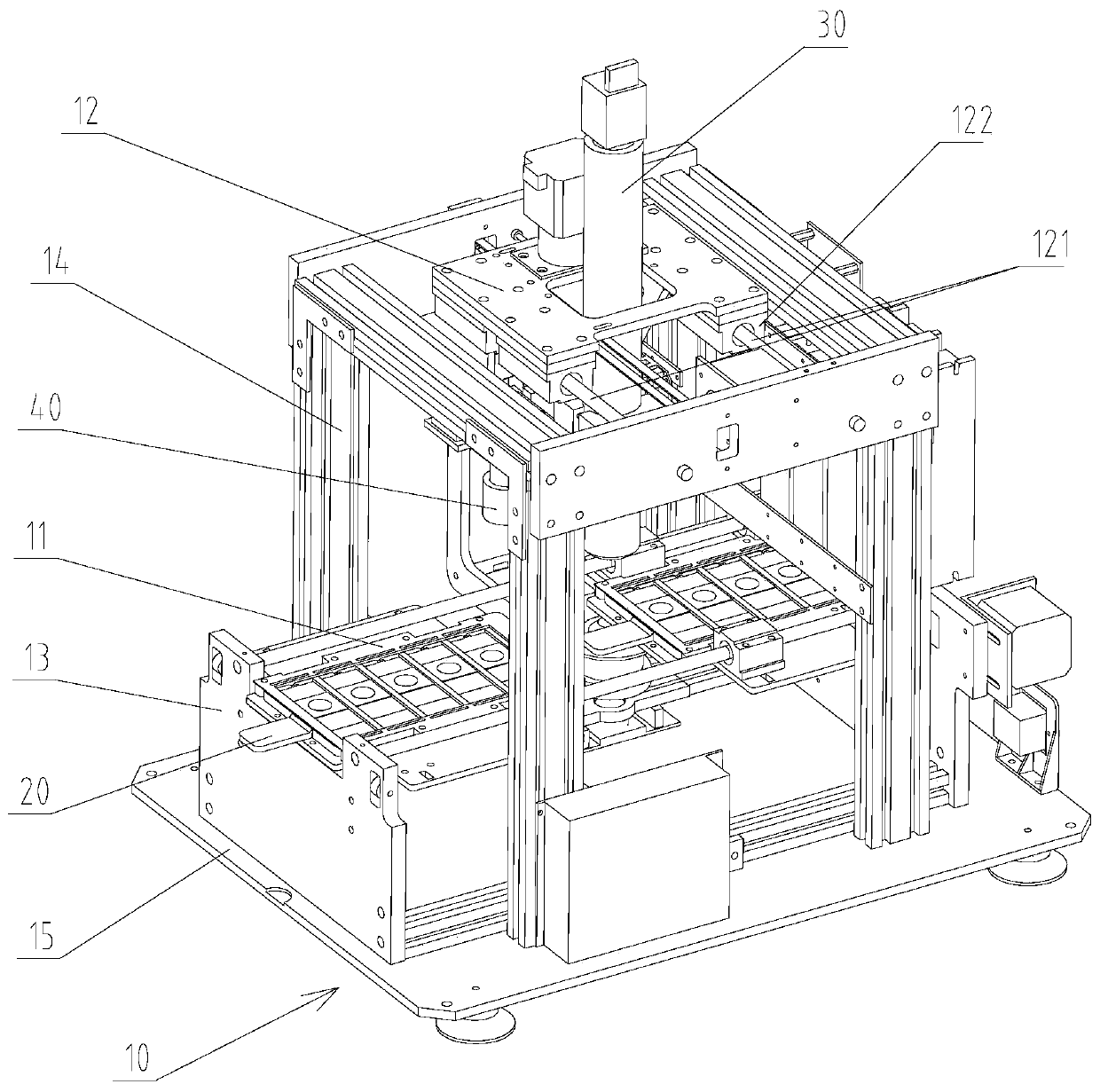
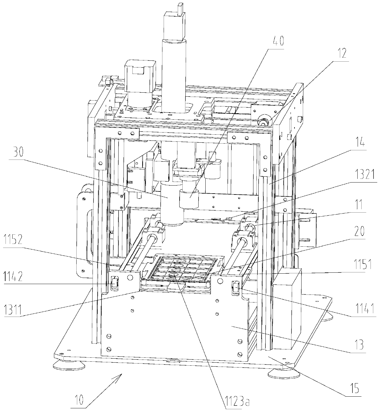
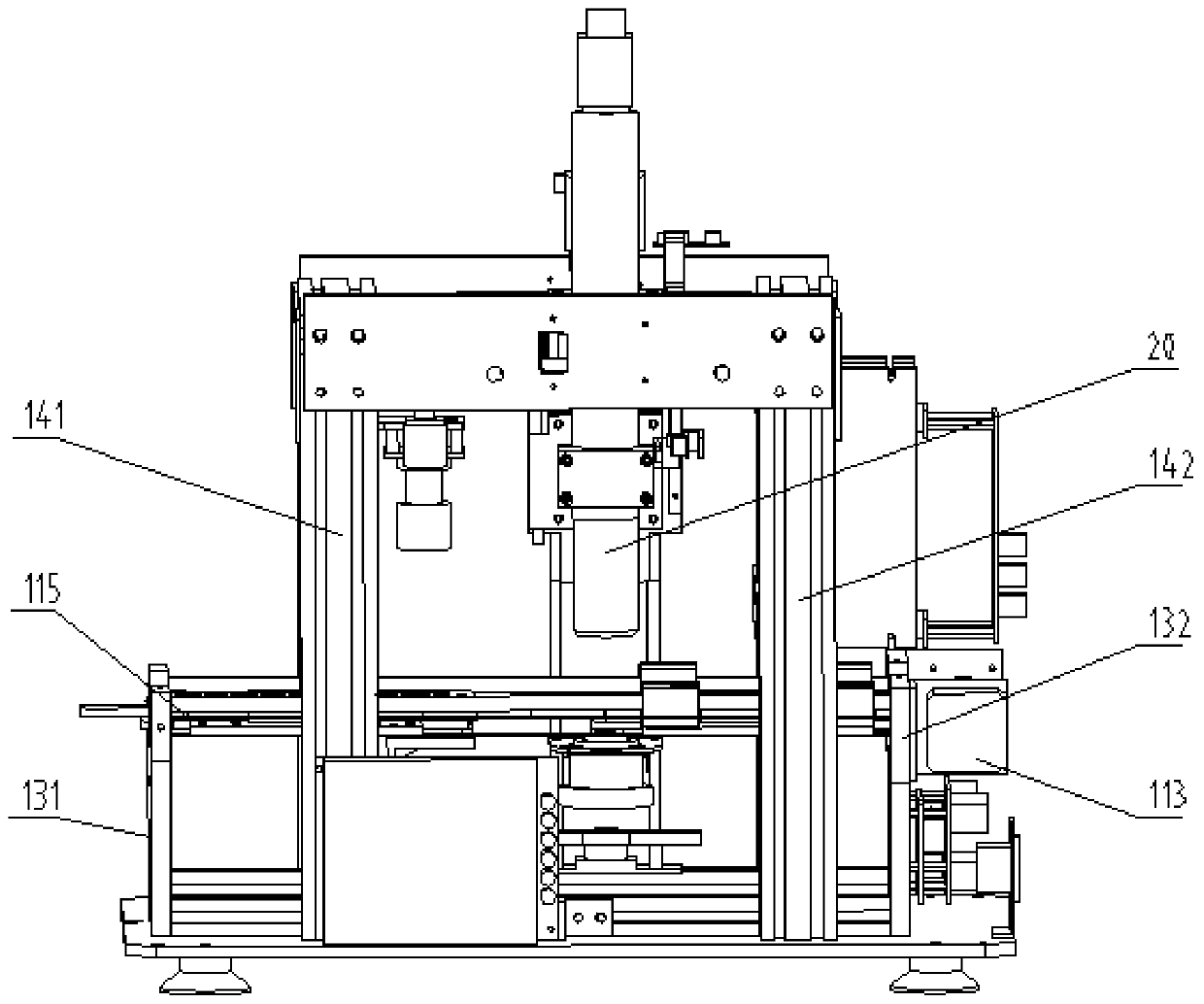
[0117] Such as Figure 1 to Figure 15 As shown, the pathological slice scanning image analysis system includes a base 10, a slide loading mechanism 20, and a slide imaging scanning mirror 30. The base 10 includes an X-axis transmission mechanism 11 and a Y-axis transmission mechanism 12. The Y-axis transmission mechanism 12 is located at above the X-axis transmission mechanism 11 , and the transmission directions of the two are perpendicular to each other; The slide loading mechanism 20 and the slide imaging scanning mirror 30 are connected to an external computer through a data line. The external computer includes a controller and a camera output device, and the external computer remotely intelligently focuses and scans the slide.

[0118] Wherein, the transmission direction of the X-axis transmission mechanism 11 is transmitted along the X-axis direction, the transmission direction of the Y-axi...


PUM


Abstract

The invention discloses a pathological section scanning image analysis system, comprising a base, a slide loading mechanism and a slide camera shooting scanning mirror, wherein the base comprises an X-axis transmission mechanism and a Y-axis transmission mechanism, the Y-axis transmission mechanism is located on the X-axis transmission mechanism, and the transmission directions of the two are perpendicular to each other; the slide loading mechanism is installed on the X-axis transmission mechanism, and the slide camera shooting scanning mirror is installed on the Y-axis transmission mechanism.The pathological section scanning image analysis system enables the slide camera shooting scanning mirror to realize up and down movement focusing of the Z axis and horizontal movement of the Y axissimultaneously, thereby shortening the movement time, the slide camera shooting scanning mirror and the X axis where a slide is loaded are effectively separated and are upper and lower stagger to eachother so as not to interfere with each other, the slide frame moves without easily shaking, the scanning height is stable, and the scanning definition is improved.

Description

technical field [0001] The invention relates to the field of medical test equipment, in particular to a scanning image analysis system for pathological slices. Background technique [0002] The existing similar devices on the market are basically slides loaded on an X-axis and Y-axis horizontal moving platform, and the camera microscope moves up and down along the Z axis to focus and scan to take pictures. Due to the reason of the camera microscope, the movement of the horizontal moving platform The guide rail can only be done on one side, so the slide loading assembly mechanism must be fixed in a cantilever type. The disadvantage of this method is that the slide rack is easy to shake, and it is easy to vibrate when moving fast, resulting in changes in the scanning height and affecting the clarity of the scan. Spend. Contents of the invention [0003] The purpose of the present invention is to provide a pathological slice scanning image analysis system, the pathological s...

Claims


Application Information

Patent Timeline
Patent Type & Authority Applications(China)
IPC IPC(8): G01N21/84G02B21/00G02B21/34G02B21/24G02B21/36
CPCG01N21/84G02B21/0012G02B21/002G02B21/34G02B21/241G02B21/362
Inventor 方陕渊陈吾科
Owner 广州英特美迪科技有限公司
Learn More