Method and device for determining scanning part and terminal equipment

A technology for scanning parts and determining methods, which can be used in computer tomography scanners, instruments for radiological diagnosis, medical science, etc., and can solve problems such as inability to determine body parts

Active Publication Date: 2019-10-11
SHENYANG INTELLIGENT NEUCLEAR MEDICAL TECH CO LTD
View PDF14 Cites 1 Cited by
  • Summary
  • Abstract
  • Description
  • Claims
  • Application Information

AI Technical Summary

Problems solved by technology

[0004] In view of this, the present application provides a method, device and terminal equipment for determining the scanning part to solve the problem in the prior art that the corresponding body parts cannot be determined using the image data of PET itself

Method used


Image

  • Method and device for determining scanning part and terminal equipment
  • Method and device for determining scanning part and terminal equipment
  • Method and device for determining scanning part and terminal equipment

Examples


Embodiment Construction

[0031] Reference will now be made in detail to the exemplary embodiments, examples of which are illustrated in the accompanying drawings. When the following description refers to the accompanying drawings, the same numerals in different drawings refer to the same or similar elements unless otherwise indicated. The implementations described in the following exemplary embodiments do not represent all implementations consistent with this application. Rather, they are merely examples of apparatuses and methods consistent with aspects of the present application as recited in the appended claims.

[0032] The terminology used in this application is for the purpose of describing particular embodiments only, and is not intended to limit the application. As used in this application and the appended claims, the singular forms "a", "the", and "the" are intended to include the plural forms as well, unless the context clearly dictates otherwise. It should also be understood that the term...


PUM


Abstract

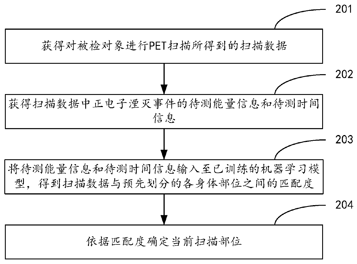
The invention provides a method and device for determining a scanning part and terminal equipment. The method comprises the steps that scanning data obtained by performing PET scanning on a detected object is obtained; to-be-measured energy information and to-be-measured time information of a positron annihilation event in the scanning data are obtained; the to-be-measured energy information and the to-be-measured time information are input to a trained machine learning model to obtain matching degrees between the scanning data and pre-divided body parts; according to the matching degrees, thecurrent scanning part is determined. The method has the advantages that the corresponding body parts can be determined by using the image data of PET itself, and the current scanning part can be determined in real time when PET scanning is performed on the detected object.

Description

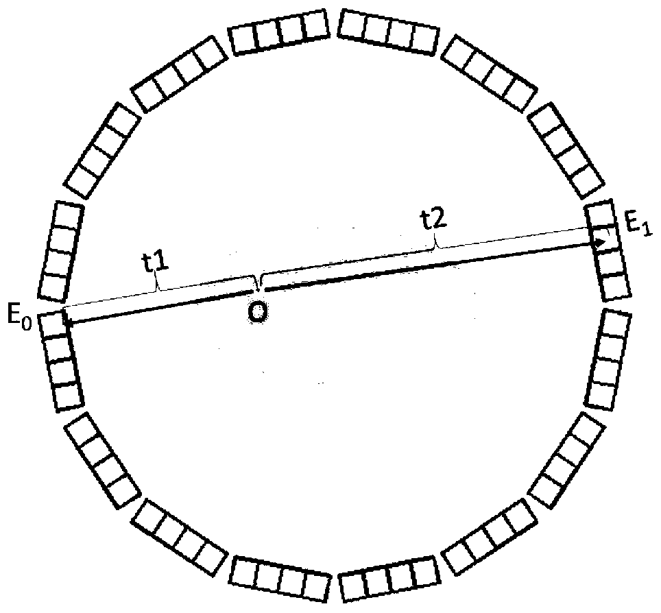
technical field [0001] The present application relates to the technical field of medical equipment, and in particular to a method, device and terminal equipment for determining a scanning part. Background technique [0002] At present, PET (Positron Emission Computed Tomography, Positron Emission Computed Tomography) is a relatively advanced clinical examination imaging technology in the field of nuclear medicine, and has been widely used in the diagnosis of various diseases and the study of organ function. When performing automatic segmentation of various organs of the human body based on PET images, blindly calling the human organ segmentation algorithm will consume a lot of Therefore, judging the body part corresponding to the currently processed PET image is the primary problem in realizing the automatic segmentation of various organs of the human body. [0003] In related technologies, existing images (for example, images from CT scans) are usually used to determine bo...

Claims


Application Information

Patent Timeline
Patent Type & Authority Applications(China)
IPC IPC(8): A61B6/03
CPCA61B6/037A61B6/5205A61B6/52Y02E30/30
Inventor 孙智鹏刘勺连李运达李明
Owner SHENYANG INTELLIGENT NEUCLEAR MEDICAL TECH CO LTD
Why Eureka
Try Eureka