Determination method and device of reconstruction data, medical imaging equipment and medium
A technology for reconstructing data and determining methods, applied in the field of biomedical image processing, can solve problems such as destroying the consistency and integrity of projection data, affecting doctors' normal diagnosis, etc., to avoid long-term dynamic scanning, reduce side effects, and avoid anesthesia effects.
- Summary
- Abstract
- Description
- Claims
- Application Information
AI Technical Summary
Problems solved by technology
Method used
Image
Examples
Embodiment 1
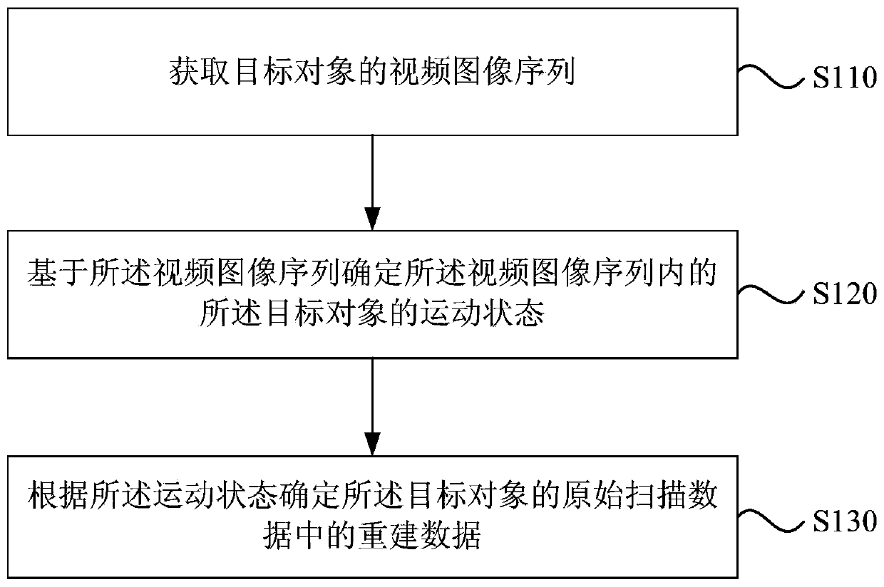
[0045] figure 1 It is a flow chart of a method for determining reconstruction data provided by Embodiment 1 of the present invention. This embodiment can be applied to obtain scan data corresponding to the time period during which the target object’s motion state is detected and confirmed to have no motion state. In the case of reconstruction, the method may be performed by a device for determining reconstruction data, and specifically includes the following steps:
[0046] S110. Acquire a video image sequence of a target object.
[0047] Wherein, the video image sequence is one or more groups
[0048] Specifically, there are two cameras on the PET side and the CT side of the 2m PET-CT equipment, and the images collected by the two cameras can complement each other in the image display content. For example, the head of the target object can be clearly seen on the PET side Whether the target object’s lower limbs are moving can be clearly seen on the CT side. The camera trans...
Embodiment 2
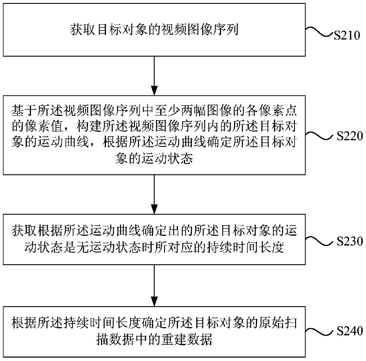
[0057] figure 2 It is a flowchart of a method for determining reconstruction data provided in Embodiment 2 of the present invention. This embodiment is optimized on the basis of the above-mentioned embodiments. In this embodiment, the step of determining the motion state of the target object in the video image sequence based on the video image sequence is further optimized as: based on the video image The pixel values of each pixel in at least two images in the sequence construct a motion curve of the target object in the video image sequence, and determine the motion state of the target object according to the motion curve. On this basis, the step of determining the reconstructed data in the original scan data of the target object according to the motion state is further optimized as follows: when the motion state of the target object determined according to the motion curve is a non-motion state The corresponding duration; determine the reconstructed data in the original...
Embodiment 3
[0073] image 3 It is a flowchart of a method for determining reconstruction data provided by Embodiment 3 of the present invention. The embodiment of the present invention is optimized on the basis of the above-mentioned embodiments. In this embodiment, the steps are to determine the video image sequence based on the video image sequence. The motion state of the target object in the image sequence is further optimized as: determining the target detection area of the target object according to the video image sequence; based on at least two images in the video image sequence and the at least two images The pixel values of each pixel in the target detection area construct a motion curve of the target object in the target detection area, and determine the motion state of the target object according to the motion curve.
[0074] Correspondingly, the method in this embodiment specifically includes:
[0075] S310. Acquire a video image sequence of the target object.
[0076] ...
PUM
 Abstract
Description
Claims
Application Information
 - R&D
- Intellectual Property
- Life Sciences
- Materials
- Tech Scout
- Unparalleled Data Quality
- Higher Quality Content
- 60% Fewer Hallucinations
Browse by: Latest US Patents, China's latest patents, Technical Efficacy Thesaurus, Application Domain, Technology Topic, Popular Technical Reports.
© 2025 PatSnap. All rights reserved.Legal|Privacy policy|Modern Slavery Act Transparency Statement|Sitemap|About US| Contact US: help@patsnap.com


