
Imaging device used for radiotherapy system

An imaging device and radiation therapy technology, applied in the medical field, can solve problems such as large differences in viewing angles, unfavorable post-processing, and visual observation by operators, and achieve the effects of convenient visual observation, radiation protection, and prevention of damage

Pending Publication Date: 2019-11-22
JIANGSU RAYER MEDICAL TECH GO LTD
View PDF0 Cites 2 Cited by
  • Summary
  • Abstract
  • Description
  • Claims
  • Application Information

AI Technical Summary

Problems solved by technology

[0006] However, the angle of view of the projected image generated under the oblique imaging geometry is quite different from that of the conventional X-ray film, which is not conducive to post-processing and visual observation by the operator.

Method used


Image

  • Imaging device used for radiotherapy system
  • Imaging device used for radiotherapy system
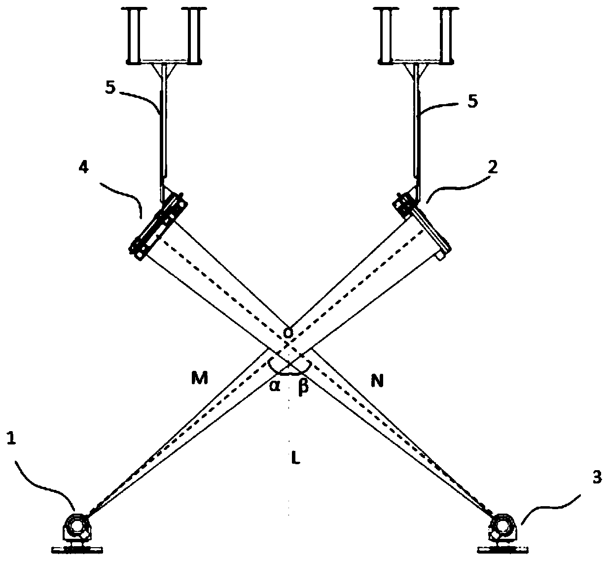
  • Imaging device used for radiotherapy system

Examples


Embodiment Construction

[0021] The specific implementation manners of the present invention will be described in detail below in conjunction with the accompanying drawings.

[0022] The present invention provides an imaging device used in a radiotherapy system, which is based on an orthogonal imaging geometry and can generate a projected image with a viewing angle similar to that of a conventional X-ray film, thereby facilitating post-processing of the image and visual observation by the operator .

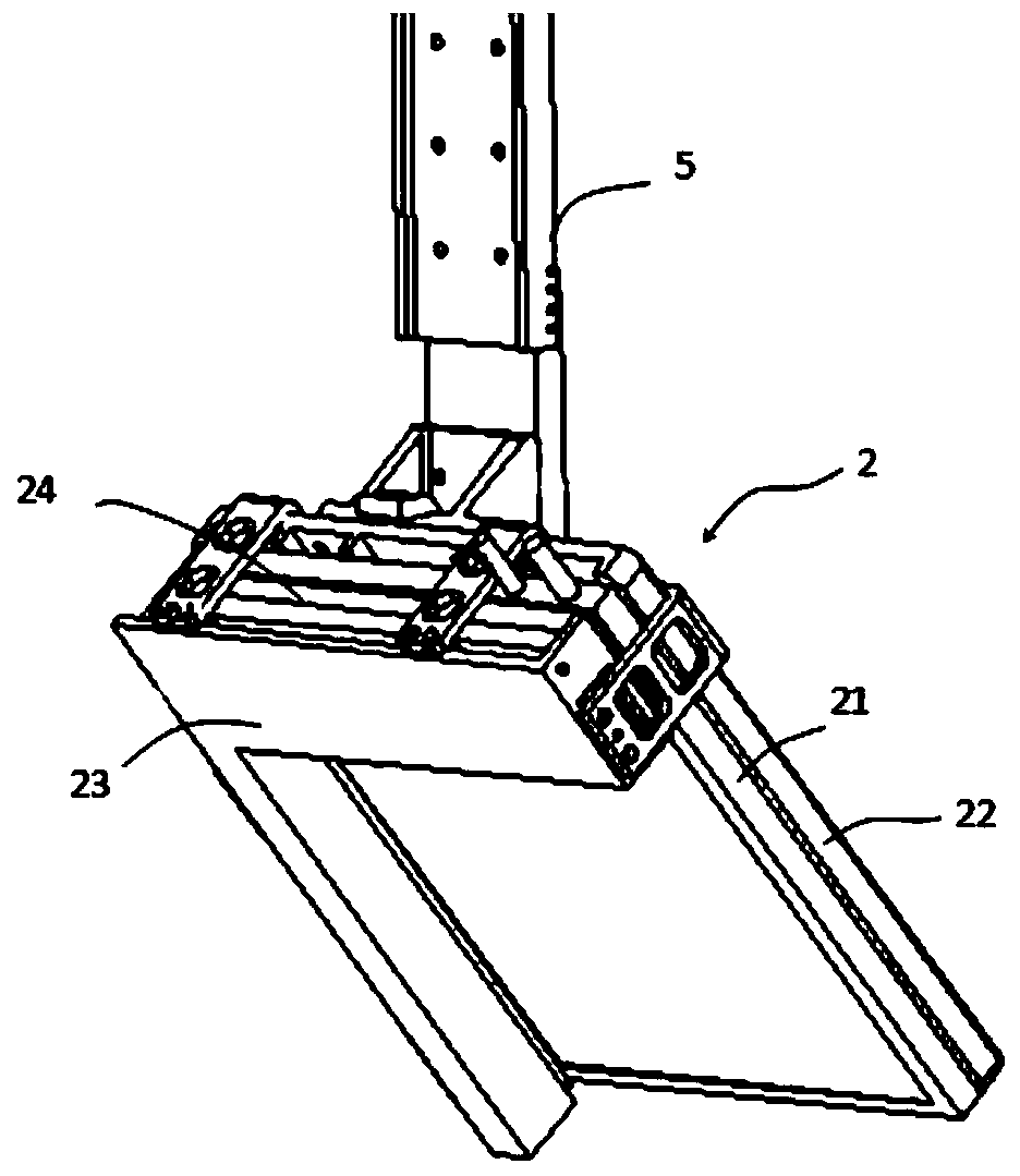
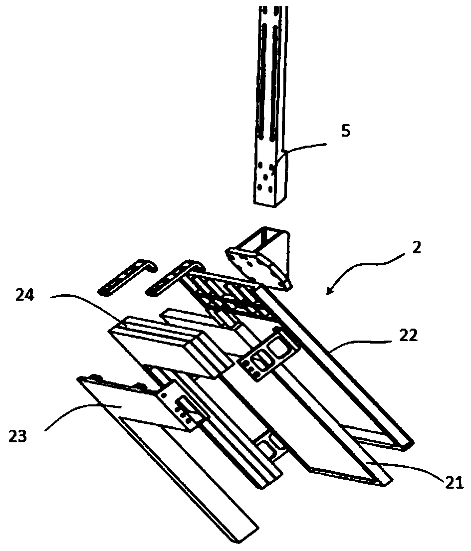
[0023] Such as figure 1 As shown, the imaging device includes a first X-ray tube 1 , a second X-ray tube 3 , a first flat panel detector 2 and a second flat panel detector 4 .

[0024] The first X-ray tube 1 is used to generate a first X-ray beam with a first optical axis M, and the second X-ray tube 3 is used to generate a second X-ray beam with a second optical axis N, and the first X-ray beam and The second X-ray beam is set to intersect at a predetermined imaging center o, and the first optical axi...


PUM


Abstract

The invention provides an imaging device used for a radiotherapy system. The imaging device comprises a first X-ray tube, a second X-ray tube, a first flat panel detector and a second flat panel detector, wherein a first X-ray wave beam generated by the first X-ray tube and a second X-ray wave beam generated by the second X-ray tube cross at the predetermined imaging center, a first optical axis of the first X-ray wave beam and a second optical axis of the second X-ray wave beam are located on the same vertical plane, and an included angle between the first optical axis and the vertical direction is equal to that between the second optical axis and the vertical direction; the first flat panel detector is used for detecting the first X-ray wave beam, and the second flat panel detector is used for detecting the second X-ray wave beam; the first flat panel detector and the second flat panel detector respectively comprise fixed frames and detector bodies installed in the fixed frames, andcircuits of the detector bodies are hidden in the fixed frames. According to the imaging device, based on orthogonal imaging geometry, projection images similar to view angles of conventional X-ray images can be generated by the imaging device, and therefore convenience is provided for image postprocessing and visual observation of operation workers.

Description

technical field [0001] The invention relates to the medical field, in particular to an imaging device used in a radiotherapy system. Background technique [0002] Image-guided Radiation Therapy (IGRT) is a new technology of tumor radiation therapy developed gradually in the past ten years. It is a milestone in the division of modern radiation therapy and has been widely used clinically at home and abroad. Image guidance uses advanced imaging equipment and image processing methods to accurately position the patient's target area before treatment and track it during treatment to improve the accuracy of radiotherapy, allow higher doses of radiation to the target area, and reduce the impact on normal tissues surrounding the tumor. And the damage of key organs, improve the treatment effect. [0003] Image-guided radiotherapy positioning system (IGPS) is one of the implementation methods of IGRT technology. It adopts kV-level X-ray stereo planar imaging technology, and obtains X-...

Claims


Application Information

Patent Timeline
IPC IPC(8): A61N5/10
CPCA61N5/1049A61N2005/1061A61N2005/1092
Inventor 马善达申志潮
Owner JIANGSU RAYER MEDICAL TECH GO LTD
Learn More