Cranial nerve automatic segmentation method based on large sample data driving

A data-driven, automatic segmentation technology, applied in the fields of neuroanatomy and medical imaging, can solve the problems of fiber tract imaging uncertainty and data errors, can not guarantee ROI, accuracy, etc., to achieve the effect of automatic segmentation

Active Publication Date: 2019-12-03
ZHEJIANG UNIV OF TECH
View PDF5 Cites 6 Cited by
  • Summary
  • Abstract
  • Description
  • Claims
  • Application Information

AI Technical Summary

Problems solved by technology

[0005] The existing imaging technology relies too much on the operation of medical workers, and has a strong subjectivity. At the same time, it cannot guarantee the accuracy of the ROI drawn each time, which may easily cause fiber tract imaging uncertainty and data errors.

Method used


Image

  • Cranial nerve automatic segmentation method based on large sample data driving
  • Cranial nerve automatic segmentation method based on large sample data driving

Examples


Embodiment Construction

[0027] The present invention will be further described below.

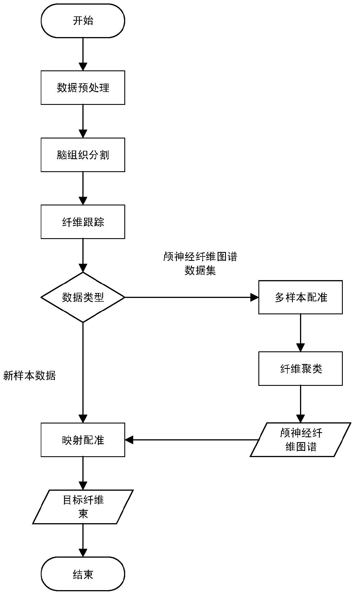
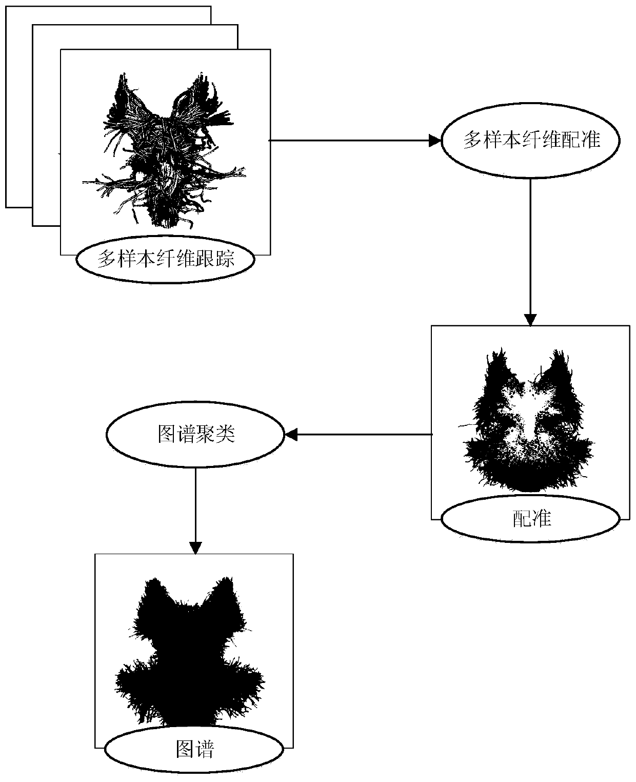
[0028] refer to figure 1 and figure 2 , an automatic segmentation method of cranial nerve fibers driven by large sample data, including the following steps:

[0029] Step 1, data preprocessing;

[0030] Each DTI data is denoised, eddy current and motion corrected to avoid some potential artifacts;

[0031] Step 2, brain tissue segmentation;

[0032] Use the recon-all command in the FreeSurfer tool to complete part or all of the FreeSurfer cortical reconstruction process. Before starting, store the structure image in a directory with a hierarchy, open the terminal under the file, enter: tcsh; enter the relevant parameters, specify A piece in the DICOM sequence, name the subject, and specify the folder where the subject is stored. After the segmentation is completed, get the segmentation result in the specified folder and convert it into a nii file;

[0033] Step Three, Fiber Tracking

[0034] Fiber tracking ...


PUM


Abstract

The invention discloses a cranial nerve fiber automatic segmentation method based on large sample data driving. A FreeSurfer tool is used for carrying out brain region segmentation. A deterministic tracking method based on spherical deconvolution (SD_STREAM) is applied to perform fiber tracking of brainstem and cerebellum parts on original DTI data, and on the basis of a cranial nerve fiber atlasobtained by a multi-sample registration and clustering method, sample data is mapped and registered to obtain a target cranial nerve fiber bundle. Compared with a common method for manually drawing ROI and removing wrong fibers, the method has the advantages that subjectivity introduced by manual operation is eliminated, meanwhile, the imaging determinacy of the fiber bundle is guaranteed, data errors are reduced, and an efficient, accurate, stable and repeatable method can be provided for cranial nerve fiber segmentation.

Description

technical field [0001] The invention relates to the fields of medical imaging and neuroanatomy under computer graphics, in particular to an automatic cranial nerve segmentation method based on fiber atlas. Background technique [0002] Cranial nerves are 12 pairs of left and right nerves that originate from the brain, and are in charge of important functions such as smell, vision, and eye movement. Among the 12 pairs of cranial nerves, except for the olfactory and hypoglossal nerves, the remaining 10 pairs of nerves are more likely to be damaged. Injury to the cranial nerves can cause loss of general sensation in the skin of the head and face and mucous membranes of the tongue, mouth, and nasal cavity, dyskinesia of the masticatory muscles on the affected side, and loss of corneal reflexes. During the treatment of cranial nerves, fiber imaging technology is used to make up for the deficiency of clinical examination in the diagnosis of pathological processes. Diffusion tens...

Claims


Application Information

Patent Timeline
Patent Type & Authority Applications(China)
IPC IPC(8): G06T7/10G06T7/11G06T7/33G06T5/00G06K9/62
CPCG06T7/10G06T7/11G06T7/33G06T5/002G06T2207/30016G06F18/23
Inventor 冯远静陈余凯金儿谭志豪曾庆润李思琦裘新宇
Owner ZHEJIANG UNIV OF TECH
Why Eureka
Try Eureka