
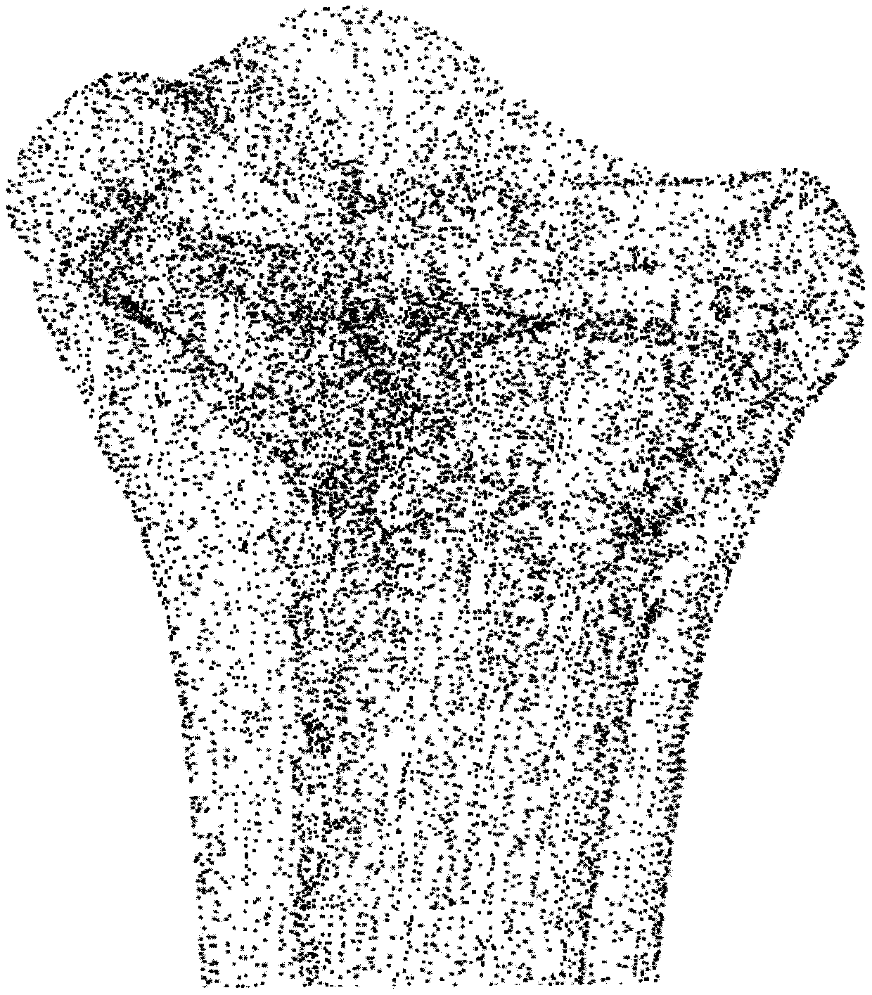
Method for carrying out human skeleton three-dimensional modeling by utilizing CT image

A CT image and three-dimensional modeling technology, applied in the field of three-dimensional modeling of human bones, can solve problems such as inability to adapt to and meet the needs of orthopedic surgery research and medical care, and inability to fully construct and reproduce the three-dimensional shape of the bones.

Pending Publication Date: 2019-12-13
TIANJIN POLYTECHNIC UNIV
View PDF0 Cites 15 Cited by
  • Summary
  • Abstract
  • Description
  • Claims
  • Application Information

AI Technical Summary

Problems solved by technology

Processing and transforming CT images in DICOM format is the basis for establishing a three-dimensional model of bones. The existing developed and commercial medical image processing software and the three-dimensional modeling method of bones cannot completely construct and reproduce the three-dimensional shape of bones. To adapt to and meet the needs of orthopedic surgery research and medical treatment in actual work, the present invention proposes a method for three-dimensional modeling of human bones using CT images

Method used


Image

  • Method for carrying out human skeleton three-dimensional modeling by utilizing CT image
  • Method for carrying out human skeleton three-dimensional modeling by utilizing CT image
  • Method for carrying out human skeleton three-dimensional modeling by utilizing CT image

Examples


Embodiment Construction

[0055] In order to further understand the invention content, characteristics and effects of the present invention, the following examples are listed, and the present invention is further described in detail in conjunction with the accompanying drawings.

[0056] (1) Completed CT scan data collection in the orthopedics department of a certain hospital, and collected full-length CT scans of the lower limbs from the tibia to the ankle joint, in which the knee joint was collected except for the straight position (0°). Male, aged 29 years, weight 75Kg, height 175cm, no Skeletal and body disorders. The slice distance of CT scanning is 1mm, and the image size is 256x256 pixels. The layer distance and resolution can meet the requirements of later image interpolation, segmentation processing, and three-dimensional reconstruction of the tibial structure, and a total of 482 images were formed. The image output format is DICOM format.

[0057] (2) Divide the single-slice CT image of the...


PUM


Abstract

The technical scheme of the invention is as follows: the method comprises the following steps: firstly, using medical CT for scanning human skeletons to obtain a DICOM format image map; increasing theresolution of the image sequence along the CT scanning direction by adopting a slice gradient interpolation method, selecting a maximum entropy segmentation method based on the probability distribution of each gray level of the image to determine a threshold parameter, and carrying out binary segmentation processing on the image according to the calculated threshold parameter; obtaining a certainnumber of discrete three-dimensional structure feature points of a two-dimensional plane of a CT scanning skeleton tomographic map, defining tetrahedral voxels of an isosurface by utilizing the distances between the three-dimensional structure feature points, generating a tetrahedral voxel set through a volume fitting method to obtain an accurate topological structure model, and generating a human skeleton three-dimensional model formed by splicing and fitting the tetrahedral voxels.

Description

technical field [0001] The invention relates to the technical field of simulation of medical images, in particular to a method for three-dimensional modeling of human skeletons using CT images. Background technique [0002] The use of CT images for bone three-dimensional modeling technology is more and more widely used. This technology does not need to dissect the human body and has no bioethical problems. The three-dimensional bone model is more and more valued by researchers and clinicians in the computer-aided diagnosis and treatment of medical departments such as orthopedics and surgery. The established three-dimensional bone model can provide a medical research platform for simulated surgical resection, joint replacement, and medical guides. Processing to help medical students understand the anatomical structure and the diagnosis of bone injuries more easily, and create surgical training opportunities for young doctors who have just entered the job. [0003] The curren...

Claims


Application Information

Patent Timeline
IPC IPC(8): G06T17/00G06T3/40G06T7/136
CPCG06T17/00G06T3/4007G06T7/136G06T2207/30008G06T2207/10081
Inventor 李雅峰张玺李毅孙杰杨涛李书震孟建军冯允佳李若琳
Owner TIANJIN POLYTECHNIC UNIV
Learn More