
Techniques for Quality Assurance of Patient Positioning Prior to Mammography Image Acquisition

A technology of images, diagnostic images, used in mammography, patient positioning for diagnosis, instruments for radiological diagnosis, etc., can solve problems such as patient burden, revocation of licenses, suboptimal imaging, etc.

Pending Publication Date: 2019-12-17
HOLOGIC INC
View PDF10 Cites 0 Cited by
  • Summary
  • Abstract
  • Description
  • Claims
  • Application Information

AI Technical Summary

Problems solved by technology

Second, it is desirable to minimize the patient's total exposure to radiation during the course of the study, so subsequent imaging in order to obtain correct positioning is not ideal
Third, due to regulations in many jurisdictions, follow-up imaging used only to correct poor positioning may be statistically targeted against the practitioner or organization, and frequent re-imaging can lead to revocation of license and / or accreditation
Fourth, incorrect positioning of images may require the patient to subsequently visit an imaging center, thereby placing an additional burden on the patient

Method used


Image

  • Techniques for Quality Assurance of Patient Positioning Prior to Mammography Image Acquisition
  • Techniques for Quality Assurance of Patient Positioning Prior to Mammography Image Acquisition
  • Techniques for Quality Assurance of Patient Positioning Prior to Mammography Image Acquisition

Examples


Embodiment Construction

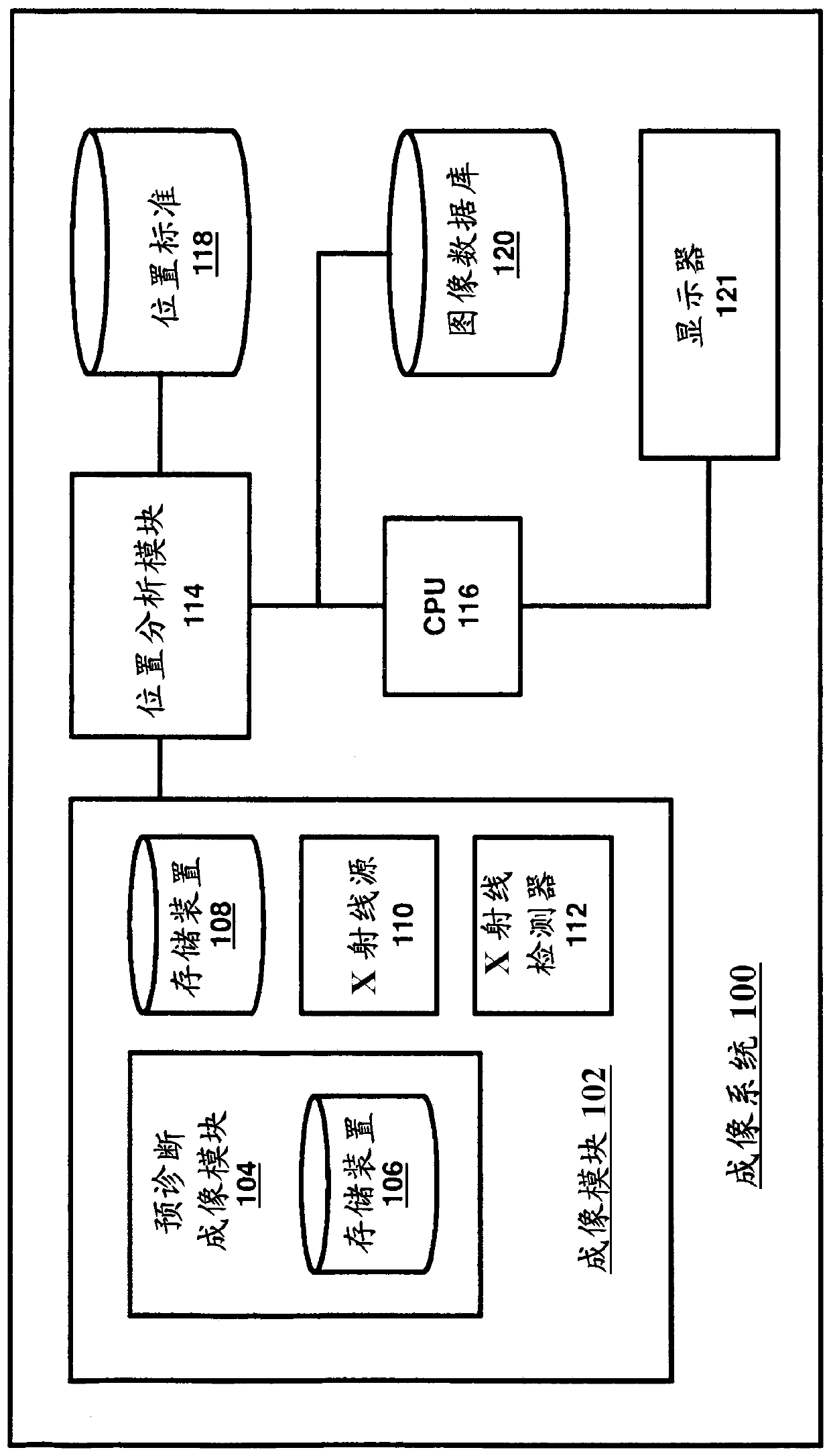
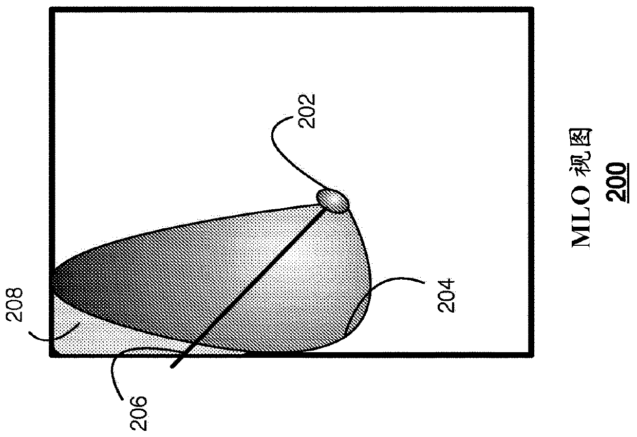
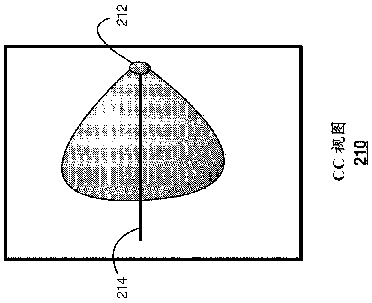
[0030] Various embodiments relate generally to describing techniques for patient positioning quality assurance prior to mammographic image acquisition. In an embodiment, the system may include an x-ray source configured to expose human tissue to radiation. X-ray detectors can be configured to detect radiation and generate pre-diagnostic images of human tissue. As used herein, a pre-diagnostic image is an image in which (a) a relatively small amount of radiation exposure to tissue is utilized and analyzed to determine generation (such as may be, but not necessarily, be controlled by an automatic exposure control of a breast imaging system) system-generated) optimal exposure parameters for diagnostic-quality images; and (b) are not intended to be presented to physicians or other healthcare providers or staff for clinical or diagnostic interpretation. Figure 12A with Figure 12B An exemplary comparison between diagnostic and pre-diagnostic images is illustrated. The position ...


PUM


Abstract

Techniques for quality assurance of patient positioning prior to mammographic image acquisition are described. The system can include an x-ray source configured to expose human tissue to radiation. The X-ray detector module can be configured to detect radiation and generate pre-diagnostic images of human tissue. The position analysis module may be configured to receive the pre-diagnostic image and determine whether the body tissue is correctly located based on one or more predefined positioning criteria. The non-transitory computer readable storage medium may be configured to store the results of the determination by the location analysis module. The display may be configured to display the location analysis module's determination within the user interface. Other embodiments are described and claimed.

Description

[0001] Cross References to Related Applications [0002] This application claims the benefit of U.S. Provisional Application No. 62 / 471,667, filed March 15, 2017, which is hereby incorporated by reference in its entirety. technical field [0003] Embodiments described herein relate generally to quality assurance of patient positioning, and more particularly to improving patient positioning in breast imaging, such as it relates to mammographic or tomosynthesis image acquisition. Background technique [0004] Proper positioning is important when performing radiation-based imaging of a patient for a number of reasons. First, the location of the target tissue is critical for obtaining high-quality images that can be used for diagnosis. Second, it is desirable to minimize the total exposure of the patient to radiation during the course of the study, so follow-up imaging to obtain correct positioning is not ideal. Third, due to regulations in many jurisdictions, follow-up imagin...

Claims


Application Information

Patent Timeline
IPC IPC(8): A61B6/04A61B6/00A61B6/02
CPCA61B6/025A61B6/04A61B6/488A61B6/502A61B6/544A61B6/542A61B6/545
Inventor B·M·凯勒井真雪
Owner HOLOGIC INC
Learn More