An adjustment structure of an ultrasonic probe rod of an extracorporeal lithotripter in the department of urology and its application method

A technology for adjusting structure and lithotripsy, applied in medical science, surgery, etc., can solve the problems of protection, poor flexibility, affecting the image quality of ultrasonic probes, etc., and achieve the effect of avoiding image quality and prolonging service life.

Active Publication Date: 2022-04-22
孙延玲
View PDF6 Cites 0 Cited by
  • Summary
  • Abstract
  • Description
  • Claims
  • Application Information

AI Technical Summary

Problems solved by technology

[0003] At present, the ultrasonic probe is moved mechanically by the lithotripter system. In clinical applications, the machine does not have thinking and is less flexible than manual operation. Especially for the final fine-tuning of the ultrasonic probe, the mechanical control needs repeated pauses. The operation is not convenient for doctors to operate, which affects the operation. In addition, the ultrasonic probe is a very precise part, which is very important for the protection of the ultrasonic probe. However, most lithotripters on the market cannot protect the ultrasonic probe when it is shut down. Shorten the service life of the ultrasound probe and affect the image quality of the ultrasound probe

Method used


Image

  • An adjustment structure of an ultrasonic probe rod of an extracorporeal lithotripter in the department of urology and its application method
  • An adjustment structure of an ultrasonic probe rod of an extracorporeal lithotripter in the department of urology and its application method
  • An adjustment structure of an ultrasonic probe rod of an extracorporeal lithotripter in the department of urology and its application method

Examples


Embodiment Construction

[0023] The following will clearly and completely describe the technical solutions in the embodiments of the present invention with reference to the accompanying drawings in the embodiments of the present invention. Obviously, the described embodiments are only some, not all, embodiments of the present invention. Based on the embodiments of the present invention, all other embodiments obtained by persons of ordinary skill in the art without making creative efforts belong to the protection scope of the present invention.

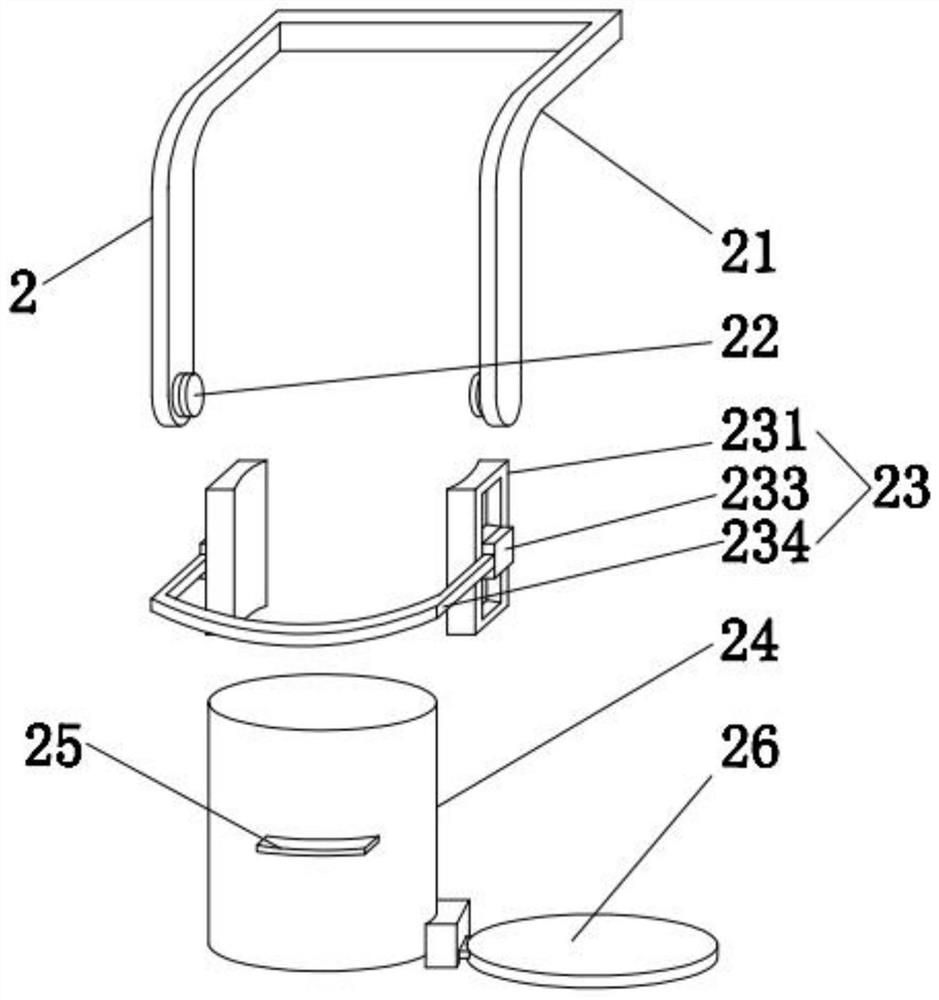
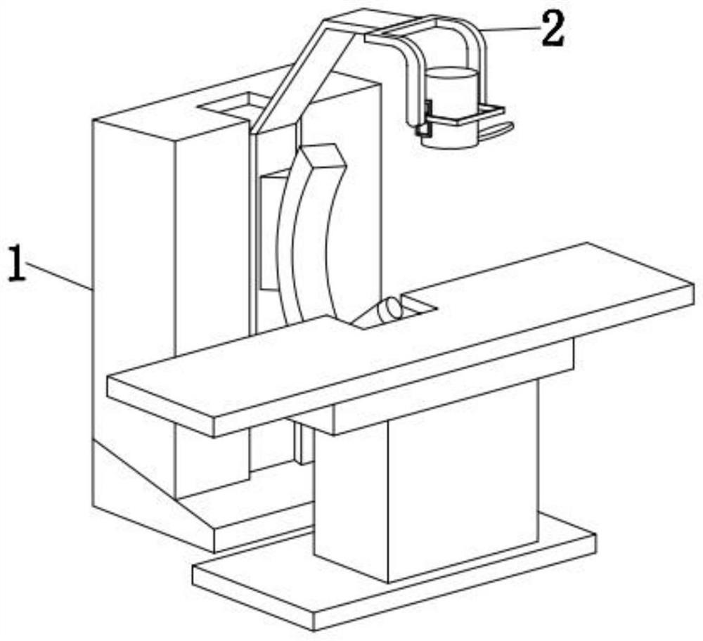
[0024] see Figure 1-4 , the present invention provides a technical solution: a urology extracorporeal lithotripter ultrasonic probe rod adjustment structure, including a lithotripter 1, the end of the mechanical arm of the lithotripter 1 is detachably equipped with an ultrasonic detection mechanism 2;

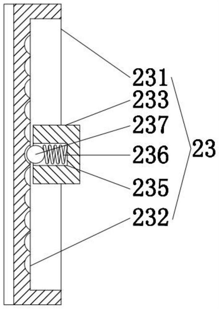
[0025] The ultrasonic detection mechanism 2 includes a bracket 21, a free stop shaft 22, a moving unit 23, an ultrasonic probe 24, a dial 25 and a sealing uni...


PUM


Abstract

The invention discloses a urology extracorporeal lithotripter ultrasonic probe rod adjustment structure and its use method, comprising a lithotripter, the mechanical arm end of the lithotripter is detachably equipped with an ultrasonic detection mechanism, and the ultrasonic detection mechanism It includes a bracket, a free-stop rotating shaft, a mobile unit, an ultrasonic probe, a dial and a sealing unit. The top of the bracket is screwed to the end of the mechanical arm of the rock crusher, and the opposite ends of the bracket are provided with a free-stop rotating shaft. The other side of the free-stop rotating shaft is screwed to the side wall of the mobile unit, and the inner side of the mobile unit is fixedly connected to the outer wall of the ultrasonic probe. In clinical use, it can realize the manual multi-directional movement of the ultrasonic probe, which is convenient for doctors to operate and facilitate the operation. In addition, it can realize the sealing of the bottom of the ultrasonic probe, protect the ultrasonic probe, prolong the service life of the ultrasonic probe, and avoid the impact Image quality of ultrasound probes.

Description

technical field [0001] The invention provides a lithotripter with a manual adjustment function, which can solve the problems in the prior art that the position of the ultrasonic probe is inconvenient to change and the protection measures are poor, which leads to the reduction of the service life of the lithotripter. Background technique [0002] The extracorporeal shock wave lithotripter usually consists of a wave source generating system, a positioning system, a water system, a three-dimensional motion system and an auxiliary system. The stone is stressed by focused high-energy pressure pulses, causing stone cracking and fragmentation. As the main part of the positioning system, the ultrasonic probe moves outside the patient's body to obtain internal images of the patient's body, so that the doctor can find the location of the stone for crushing; [0003] At present, the ultrasonic probe is moved mechanically by the lithotripter system. In clinical applications, the machin...

Claims


Application Information

Patent Timeline
IPC IPC(8): A61B17/225
CPCA61B17/2256
Inventor 孙延玲牛敏孙玲玲
Owner 孙延玲
Why Eureka
Try Eureka