Immunohistochemical karyoplasm staining section diagnosis method and system

A technique of immunohistochemistry and diagnostic method, which is applied in the field of immunohistochemical nuclear plasma staining section diagnosis method and system, and achieves the effects of small size, saving storage cost, and convenient portability

Pending Publication Date: 2020-01-31
杭州迪英加科技有限公司
View PDF6 Cites 14 Cited by
  • Summary
  • Abstract
  • Description
  • Claims
  • Application Information

AI Technical Summary

Problems solved by technology

[0003] The purpose of the present invention is to solve the problem existing in the prior art that it is necessary to scan the immunohistochemical nucleoplasmic stained section into a full-field image under a high-magnification microscope, and then upload it to a diagnostic tool for analysis. The present invention provides an immunohistochemical Diagnosis method and system for histochemical nucleoplasmic staining section

Method used


Image

  • Immunohistochemical karyoplasm staining section diagnosis method and system
  • Immunohistochemical karyoplasm staining section diagnosis method and system
  • Immunohistochemical karyoplasm staining section diagnosis method and system

Examples


Embodiment Construction

[0036] The present invention will be further described below in conjunction with accompanying drawing:
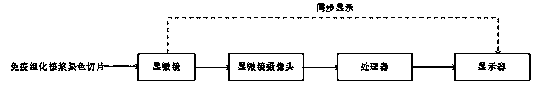
[0037] The MPP values ​​of the digital pathological full-field images used in this embodiment are 0.48, 0.96, 1.92, and 3.84 respectively. The MPP values ​​of images collected by scanners produced by different manufacturers are slightly different at the same magnification, but it does not affect The accuracy of the diagnosis results using the method provided in this application. MPP is mircons per pixel, which is a general parameter describing the image magnification, representing the length of one pixel on the screen corresponding to the physical world. The microscope used is an in-line microscope, including 1 eyepiece and 4 objective lenses; the magnification of the eyepiece is 10 times, the magnification of the objective lens is 4 times, 10 times, 20 times, 40 times, the images collected by the microscope camera The MPP values ​​are 1.5, 0.6, 0.3 and 0.15, respectively....


PUM

PropertyMeasurementUnit
diameteraaaaaaaaaa
diameteraaaaaaaaaa

Abstract

The invention relates to an immunohistochemical karyoplasm staining section diagnosis method. The method includes: S10, collecting a pathological image of the immunohistochemical karyoplasm staining section under the microscope by a microscope camera, and sending the same to a processor; S20, preprocessing the pathological image by the processor to enable HSV color space of the pathological imageto be consistent with set thresholds; S30, carrying out cell detection and cell classification on the pre-processed pathological image by the processor, labeling cell positions, cell types and corresponding immunohistochemical indexes on the pathological image, and outputting the labeled pathological image; and S40, receiving and displaying the labeled pathological images from the processor by a displayer. The application also provides an immunohistochemical karyoplasm staining section diagnosis system. By adopting the method, analysis can be realized without the need for storing a large number of GB-level digital pathological sections, and a storage overhead of the processor is saved.

Description

technical field [0001] The invention relates to the field of diagnosis of immunohistochemical nuclear and plasma stained sections, in particular to a diagnostic method and system for immunohistochemical nuclear and plasma stained sections. Background technique [0002] Applying the principles of immunology and histochemistry to conduct in situ qualitative, localization or quantitative research on certain chemical components in tissue sections or cell specimens, this technique is called immunohistochemistry or immunocytochemistry. Immunohistochemical staining is based on the combination of antigen and antibody to label specific cells to detect the changes of specific cells, so as to realize the positioning, quantification and qualitative functions of tumors. The traditional method of calculating immunohistochemical indicators is that pathologists count the percentage of positive tumor cells in the tumor area under the microscope, but the workload is huge and the accuracy is l...

Claims


Application Information

Patent Timeline
Patent Type & Authority Applications(China)
IPC IPC(8): G01N21/84G01N33/53G06T7/00G06K9/62
CPCG01N21/84G01N33/53G06T7/0012G06T2207/10024G06T2207/20081G06T2207/20084G06T2207/30024G06T2207/30096G06F18/241
Inventor 崔灿林明珍石永华沈珏伟徐建红杨林
Owner 杭州迪英加科技有限公司
Why Eureka
Try Eureka