Medical image annotation method and device, equipment and medium

A medical image and image technology, which is applied in the field of data processing, can solve the problems of inability to ensure the accuracy of the lesion and accurate location of the lesion, and inability to avoid false positives.

Inactive Publication Date: 2020-04-24
INFERVISION MEDICAL TECH CO LTD
View PDF7 Cites 3 Cited by
  • Summary
  • Abstract
  • Description
  • Claims
  • Application Information

AI Technical Summary

Problems solved by technology

However, this method cannot avoid the occurrence of false positives during the labeling process, and cannot guarantee the accuracy of the marked lesions and the accurate location of the lesions

Method used


Image

  • Medical image annotation method and device, equipment and medium
  • Medical image annotation method and device, equipment and medium
  • Medical image annotation method and device, equipment and medium

Examples


Embodiment 1

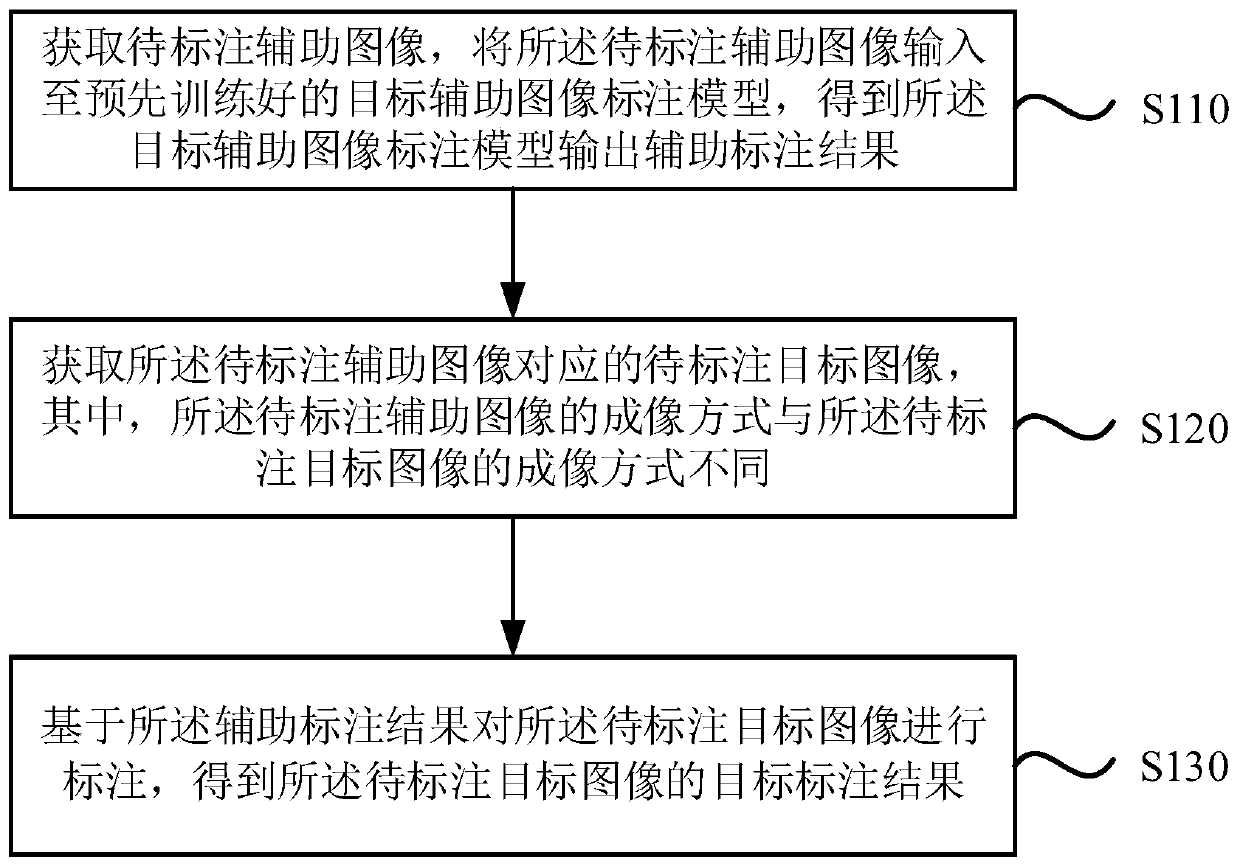
[0026] figure 1 It is a flowchart of a medical image labeling method provided by Embodiment 1 of the present invention. This embodiment is applicable to the situation of labeling medical images. The method can be executed by a medical image labeling device, which can be implemented in software and / or hardware, for example, the medical image labeling device can be configured in a computer device. like figure 1 As shown, the method includes:

[0027] S110. Acquire an auxiliary image to be labeled, input the auxiliary image to be labeled into a pre-trained object auxiliary image annotation model, and obtain an auxiliary annotation result output by the object auxiliary image annotation model.

[0028] In this embodiment, the gold standard labeling result of the auxiliary image is obtained by labeling the auxiliary image, and the target image is labeled according to the gold standard labeling result of the auxiliary image to obtain an accurately labeled target image.

[0029] O...

Embodiment 2

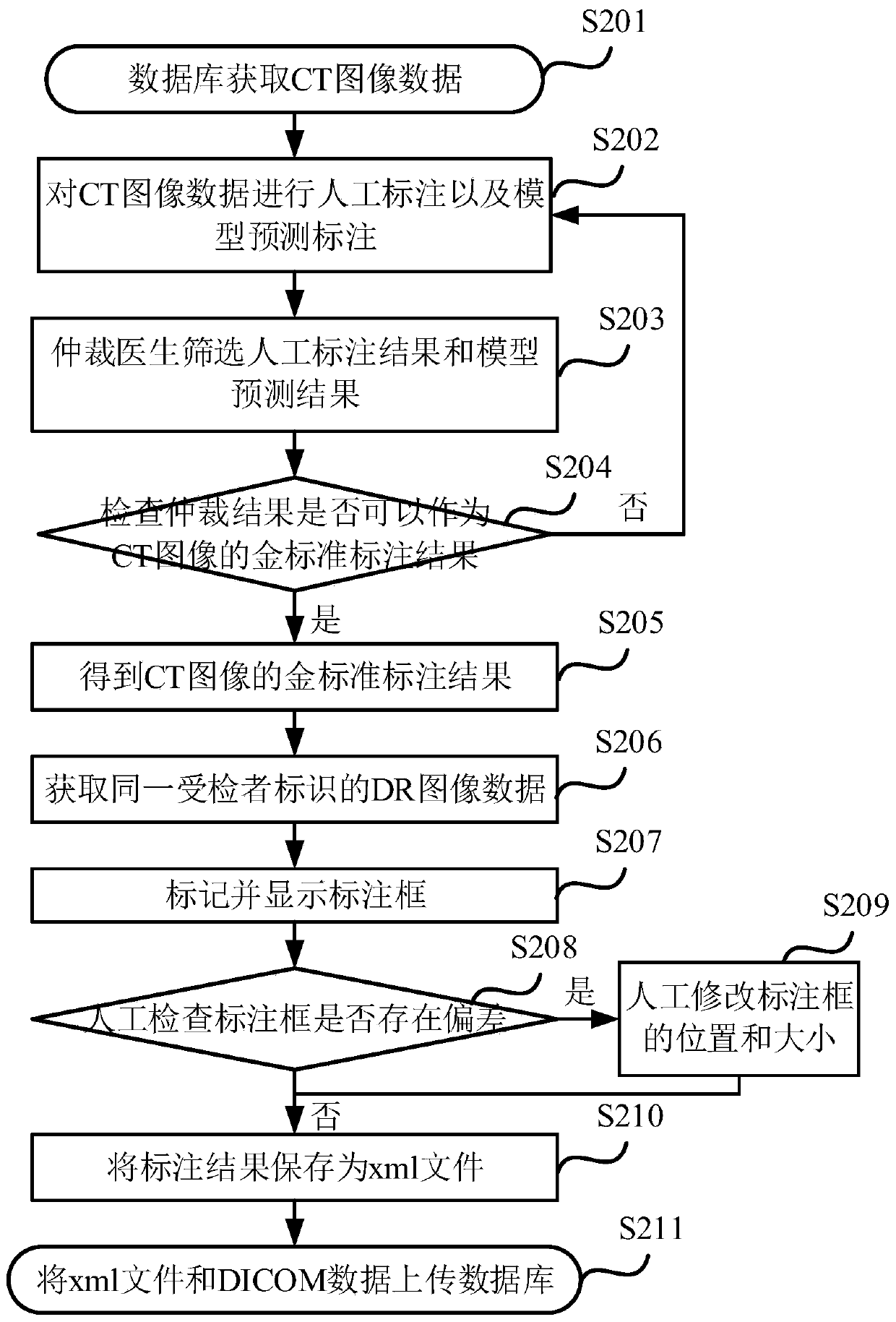
[0047] Figure 2a It is a flowchart of a medical image labeling method provided by Embodiment 2 of the present invention. This embodiment provides a preferred embodiment on the basis of the foregoing embodiments. like Figure 2a As shown, the method includes:

[0048] S201. The database acquires CT image data.

[0049] S202 , performing manual labeling and model prediction labeling on the CT image data.

[0050] First, obtain the CT image data that needs to be labeled from the database, and then assign the same CT image data to professional doctors and prediction models for labeling, and obtain manual labeling results and model prediction labeling results.

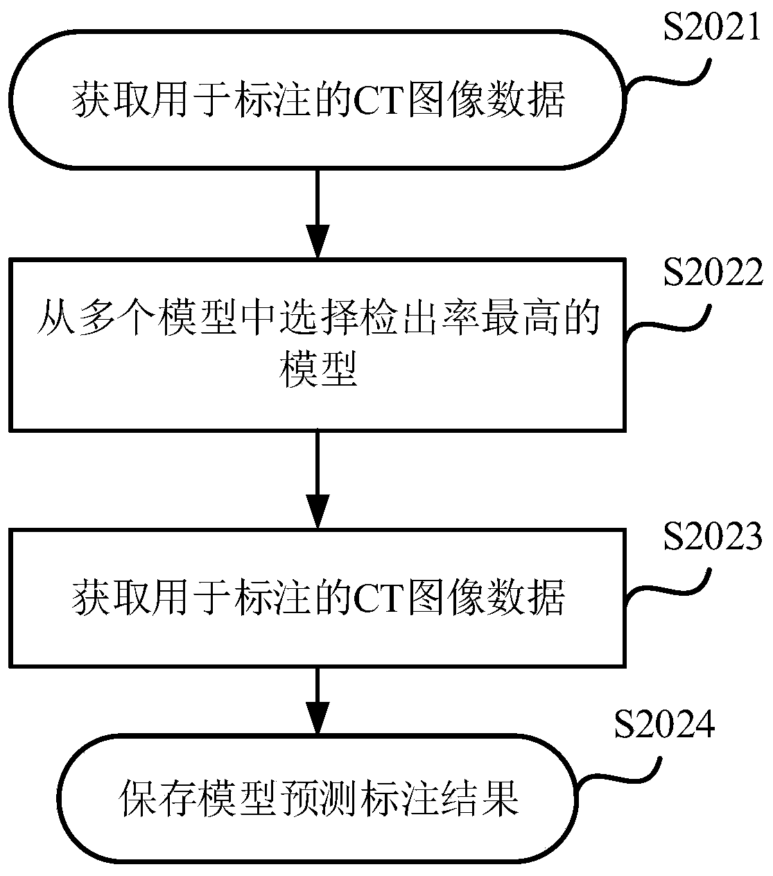
[0051] Figure 2b It is a schematic diagram of a model prediction labeling process provided by Embodiment 2 of the present invention. like Figure 2b As shown, the method includes:

[0052] S2021. Acquire CT image data for labeling.

[0053] S2022. Select the model with the highest detection rate from the multiple...

Embodiment 3

[0077] image 3 It is a schematic structural diagram of a medical image labeling device provided by Embodiment 3 of the present invention. The medical image labeling device can be realized by software and / or hardware, for example, the medical image labeling device can be configured in a computer device. like image 3 As shown, the device includes an auxiliary annotation acquisition module 310, a target image acquisition module 320 and a target annotation determination module 330, wherein:

[0078] Auxiliary annotation acquisition module 310, configured to acquire an auxiliary image to be annotated, input the auxiliary image to be annotated into a pre-trained auxiliary image annotation model for an object, obtain an auxiliary annotated result output by the auxiliary image annotated model for the target;

[0079] A target image acquisition module 320, configured to acquire a target image to be marked corresponding to the auxiliary image to be marked, wherein the imaging method o...


PUM


Abstract

The embodiment of the invention discloses a medical image annotation method and device, equipment and a medium. The method comprises the steps of obtaining a to-be-annotated auxiliary image, inputtingthe to-be-annotated auxiliary image to a pre-trained target auxiliary image annotation model, obtaining an auxiliary annotation result output by the target auxiliary image annotation model; obtaininga to-be-annotated target image corresponding to the to-be-annotated auxiliary image, wherein the imaging mode of the to-be-annotated auxiliary image is different from the imaging mode of the to-be-annotated target image; and annotating the to-be-annotated target image based on the auxiliary annotation result to obtain a target annotation result of the to-be-annotated target image. According to the medical image annotation method provided by the embodiment of the invention, the to-be-annotated target image is annotated according to the auxiliary annotation structure of the to-be-annotated auxiliary image, so that the annotation accuracy of the target image is improved.

Description

technical field [0001] The embodiments of the present invention relate to the field of data processing, and in particular to a medical image labeling method, device, equipment and medium. Background technique [0002] With the development of artificial intelligence technology, medical workers can make auxiliary diagnosis based on artificial intelligence model and improve the accuracy of clinical diagnosis. [0003] A large amount of data and data labeling are required for auxiliary diagnosis based on artificial intelligence models. Data plays a crucial role in model training of artificial intelligence. High-quality data can improve the prediction accuracy of the model. When the quality of data labeling is low, the model will be difficult to learn. In the existing data labeling methods, the purpose of labeling lesions is achieved through two labeling methods: computer tomography (Computed Tomography, CT) data labeling and direct digital radiography (Direct Digit Radiography,...

Claims


Application Information

Patent Timeline
Patent Type & Authority Applications(China)
IPC IPC(8): G16H30/20
CPCG16H30/20
Inventor 唐晨阳刘丰恺李新阳陈宽王少康
Owner INFERVISION MEDICAL TECH CO LTD
Why Eureka
Try Eureka