
Method for promoting directional differentiation of umbilical cord mesenchymal stem cells into chondrogenesis

A technology for stem cells and cartilage formation, applied in the field of stem cells and regenerative medicine, can solve the problems of waste of cell resources, high cost, long induction time, etc.

Active Publication Date: 2020-05-01
GUANGDONG VITALIFE BIOTECHNOLOGY CO LTD
View PDF4 Cites 5 Cited by
  • Summary
  • Abstract
  • Description
  • Claims
  • Application Information

AI Technical Summary

Problems solved by technology

[0005] The deficiencies of the above existing stem cell chondrogenic differentiation methods are as follows: the required induction time is longer; the use of support media to assist chondrogenic differentiation is more complicated and the cost is higher; although the cell viability will be detected before each induction, it cannot be guaranteed A batch of cells can complete chondrogenic differentiation, which will cause waste of cell resources

Method used


Image

  • Method for promoting directional differentiation of umbilical cord mesenchymal stem cells into chondrogenesis
  • Method for promoting directional differentiation of umbilical cord mesenchymal stem cells into chondrogenesis
  • Method for promoting directional differentiation of umbilical cord mesenchymal stem cells into chondrogenesis

Examples


Embodiment

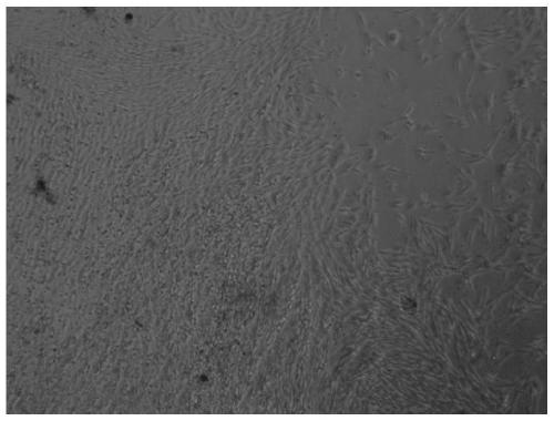
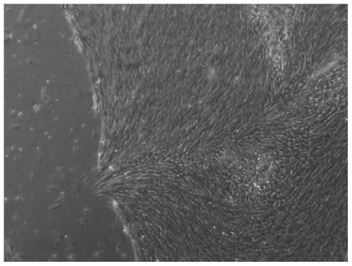
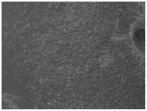
[0063] Examples 1-3 were isolated, cultured, subcultured, frozen and induced to form cartilage according to the above method. The results are shown in Table 1.

[0064] Table 1

[0065] Example 1 Example 2 Example 3 Isolate and culture for 13 days figure 1 figure 2 image 3 Cell count after isolation and culture for 13 days 5*10 6 indivual

[0066] in, Figure 1-3 It is a photo of the umbilical cord mesenchymal stem cells isolated and purified from tissue in Example 1-3 for 13 days, showing that the cells are dense and grow well; Figure 4-6 It is the photo of chondrogenic staining obtained after applying the methods of separation, cryopreservation, recovery and induction in Example 1-3, and the glycosaminoglycans of the cells are stained with toluidine blue, showing dark blue precipitates. It can be seen that all of Examples 1-3 effectively promote chondrogenic differentiation of umbilical cord mesenchymal stem cells.


PUM


Abstract

The present invention provides a method for promoting directional differentiation of umbilical cord mesenchymal stem cells into chondrogenesis. The method comprises the following steps: taking out umbilical cord tissues and conducting shredding and washing, adding tissue digestion fluid for digestion, and conducting filtering to remove supernatant after the digestion to obtain umbilical cord mesenchymal stem cells; culturing the umbilical cord mesenchymal stem cells with an umbilical cord mesenchymal stem cell selective culture medium; when the cells reach 80-90% confluence, digesting the cells with low-toxic digestion fluid, then using an umbilical cord mesenchymal stem cell cryopreservation solution for cryopreservation and placing the cells in liquid nitrogen; removing the cells from the liquid nitrogen, conducting quick thawing in a 37-DEG C water bath kettle, conducting centrifugation to remove supernatant, and adding a cell recovery solution to recover the cells; removing the recovery solution and replacing an exciting solution before induction; and when the cells grow to 80-90% confluence, digestign the cells with the low-toxic digestion solution; inoculating the cells, conducting culture overnight, removing the supernatant, adding a chondrogenesis induction culture medium, and conducting culture in a hypoxic incubator at 2% oxygen concentration. Compared with the priorart, the method can stably and effectively promote the directional differentiation of the umbilical cord mesenchymal stem cells into the chondrogenesis with shorter induction time.

Description

technical field [0001] The invention belongs to the field of stem cells and regenerative medicine, in particular to a method and culture medium for promoting chondrogenic differentiation of umbilical cord mesenchymal stem cells. Background technique [0002] Umbilical cord mesenchymal stem cells have multilineage differentiation ability. The differentiation effect of mesenchymal stem cells is mainly determined by the activity of stem cells and induction conditions. [0003] Mesenchymal stem cell chondrogenesis is a multi-step cellular event, which begins with the aggregation of a special subset of mesenchymal stem cells and then differentiates into cartilage. [0004] The commonly used method for obtaining umbilical cord mesenchymal stem cells is as follows: cut Wharton's jelly into pieces, and adhere to culture in complete medium (DMEM with 10% FBS) for 16 days. The conventional cryopreservation method is as follows: freeze in liquid nitrogen after programmed cooling in c...

Claims


Application Information

Patent Timeline
IPC IPC(8): C12N5/077A01N1/02
CPCC12N5/0655A01N1/021C12N2509/00C12N2506/1369C12N2500/02
Inventor 孙灿兴李静静赵蓝刘小翠
Owner GUANGDONG VITALIFE BIOTECHNOLOGY CO LTD
Learn More