A method and system for automatic detection of lesions in pathological tissue slice images

A lesion area and image technology, which is applied in the field of medical pathological image processing, can solve the problems of inconsistent identification results of a single lesion area, and achieve the effects of expanding the receptive field range, improving segmentation accuracy, and fast speed

Active Publication Date: 2022-07-05
HUAZHONG UNIV OF SCI & TECH
View PDF4 Cites 0 Cited by
  • Summary
  • Abstract
  • Description
  • Claims
  • Application Information

AI Technical Summary

Problems solved by technology

[0004] In view of the above defects or improvement needs of the prior art, the present invention provides a method and system for automatic detection of lesion areas in pathological tissue slice images, thereby solving the problem of a single lesion area in the existing detection of pathological images based on deep convolutional neural networks Identify technical issues with inconsistent results

Method used


Image

  • A method and system for automatic detection of lesions in pathological tissue slice images
  • A method and system for automatic detection of lesions in pathological tissue slice images
  • A method and system for automatic detection of lesions in pathological tissue slice images

Examples


Embodiment Construction

[0040] In order to make the objectives, technical solutions and advantages of the present invention clearer, the present invention will be further described in detail below with reference to the accompanying drawings and embodiments. It should be understood that the specific embodiments described herein are only used to explain the present invention, but not to limit the present invention. In addition, the technical features involved in the various embodiments of the present invention described below can be combined with each other as long as they do not conflict with each other.

[0041] The terms "first", "second", "third" and "fourth" in the description and claims of the present invention are used to distinguish different objects, rather than to describe a specific order.

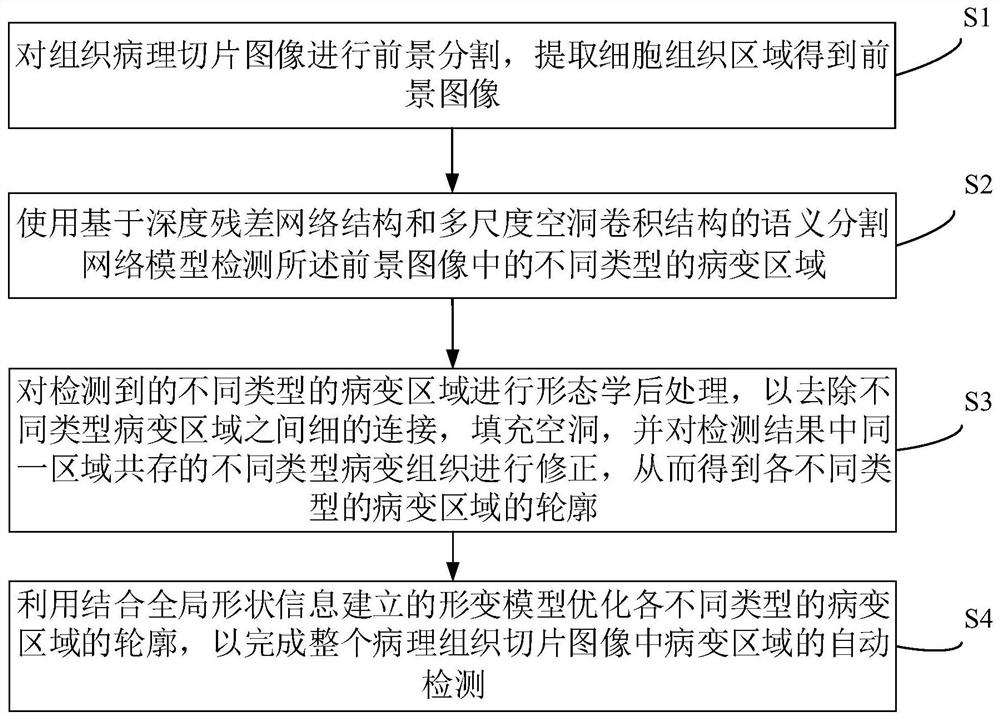
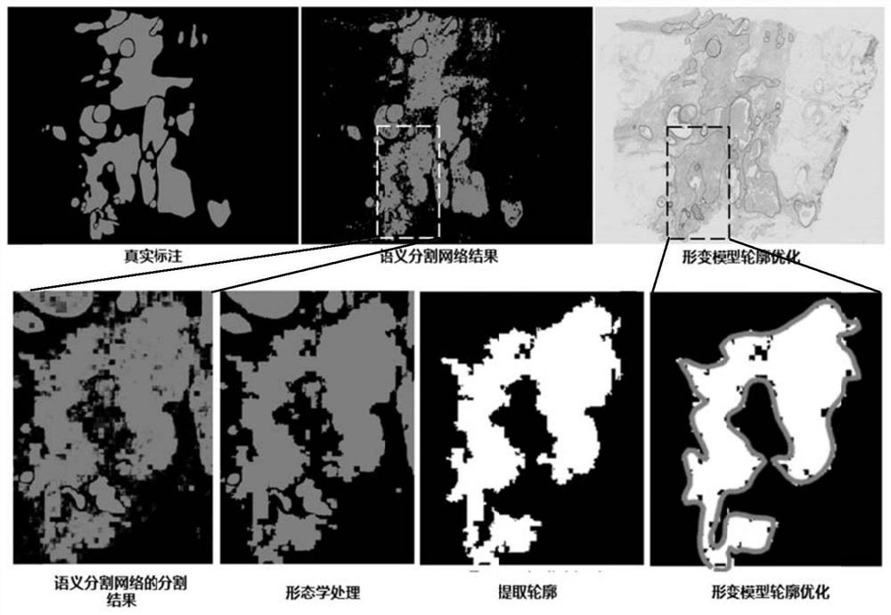
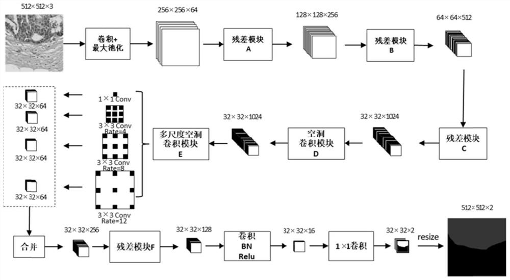
[0042] The present invention provides a method and system for automatic detection of lesion areas in histopathological slices based on a deep semantic segmentation network and a deformation model, which ...


PUM


Abstract

The invention discloses an automatic detection method and system for a lesion area in a histopathological slice image, wherein the realization of the method includes: performing foreground segmentation on the histopathological slice image, extracting a cell tissue area to obtain a foreground image; The semantic segmentation network model of structure and multi-scale hole convolution structure detects different types of lesion areas in the foreground image; morphological post-processing is performed on the detected lesion areas of different types to remove fine details between different types of lesion areas. The contours of different types of lesion areas are obtained by connecting and filling the holes; the contours of different types of lesion areas are optimized by using the deformation model established by combining the global shape information to complete the automatic detection of lesion areas in the entire pathological tissue slice image. . The combination of the deep semantic segmentation network and the deformation model proposed by the present invention can integrate prior knowledge and global information to improve the accuracy of semantic segmentation.

Description

technical field [0001] The invention belongs to the field of medical pathological image processing, and more particularly, relates to a method and system for automatic detection of lesion areas in pathological tissue slice images based on a deep semantic segmentation network and a deformation model. Background technique [0002] Breast cancer is one of the most common malignant tumors in women, and the incidence of malignant tumors in women is extremely high. Clinically, early screening for breast cancer usually involves palpation and periodic examinations, with initial detection using mammography or ultrasound imaging, followed by a breast biopsy if the detection shows the possibility of malignant tissue growth. Pathologists differentiate between normal tissue, benign and malignant lesions and perform prognostic assessments based on breast pathology images. Accurate detection and classification of pathological images is an important basis for pathologists to make the best ...

Claims


Application Information

Patent Timeline
Patent Type & Authority Patents(China)
IPC IPC(8): G06T7/00G06T7/90G06T7/194
CPCG06T7/0012G06T7/194G06T7/90G06T2207/10081G06T2207/20081G06T2207/30068
Inventor 程胜华曾绍群贾园园刘小茂
Owner HUAZHONG UNIV OF SCI & TECH
Why Eureka
Try Eureka