Three-dimensional lung feature extraction method based on CT image

A CT image and feature extraction technology, applied in the field of medical image processing, can solve the problems of insignificant feature extraction, inaccurate extraction of anatomical structures or geometric structures, and increased burden of image registration, so as to avoid the problem of poor feature point set registration. Effect

Active Publication Date: 2020-05-12
NANJING UNIV OF SCI & TECH
View PDF2 Cites 6 Cited by
  • Summary
  • Abstract
  • Description
  • Claims
  • Application Information

AI Technical Summary

Problems solved by technology

In the extraction of feature points, there are problems such as too little extraction, unremarkable features, inaccurate extracti

Method used


Image

  • Three-dimensional lung feature extraction method based on CT image
  • Three-dimensional lung feature extraction method based on CT image
  • Three-dimensional lung feature extraction method based on CT image

Examples


Embodiment

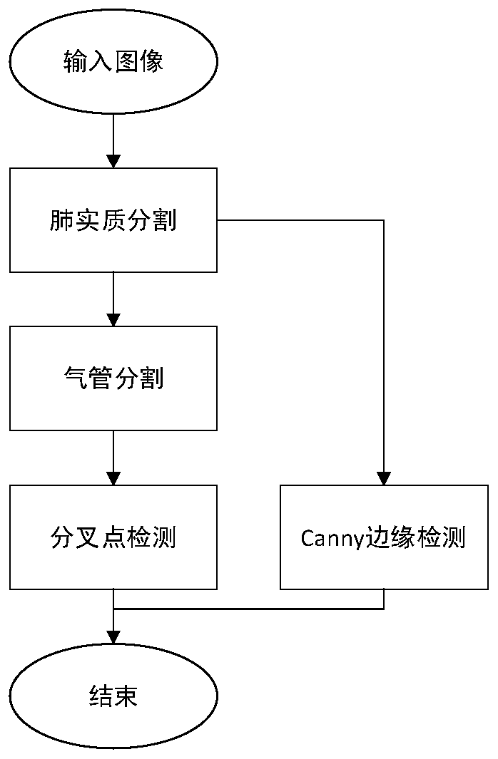
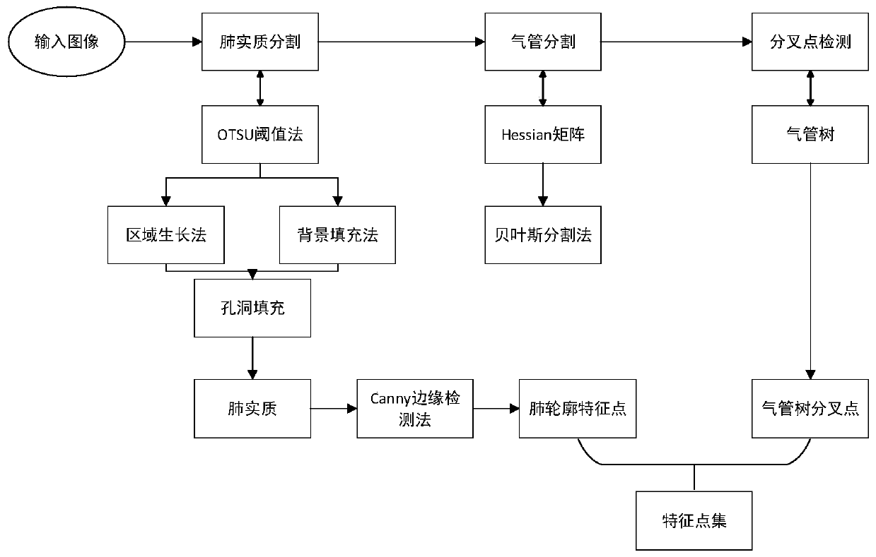
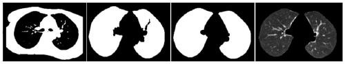
[0051] Carry out lung parenchyma segmentation on lung CT images, such as image 3 As shown, the specific steps are:

[0052] Step 101, perform binarization on the lung CT image to obtain a binarized image, in some embodiments, obtain a binarized image through OTSU algorithm (Otsu Threshold Segmentation);

[0053] Step 102, set the pixel with the gray value of the boundary line of the single binarized image as 0 to 100; traverse the pixels of the entire image from the upper left corner and the lower right corner of the image at the same time, if the four neighbors of pixel A If there is a pixel with a gray value of 0, set the gray value of pixel A to 100; re-traverse the new image, and set the gray value of the pixel with a gray value of 100 to 255 to complete the background filling;

[0054] Step 103, set the seed point for the new image, and extract the left and right lung masks by using the region growing method;

[0055] Step 104, filling the holes in the lung mask;

[0...


PUM


Abstract

The invention discloses a three-dimensional lung feature extraction method based on a CT image. The method comprises the steps of performing lung segmentation on a lung CT image to obtain lung parenchyma; performing tracheal segmentation on the pulmonary parenchyma to obtain a tracheal tree; performing bifurcation point detection on the tracheal tree to obtain a bifurcation feature point set; extracting contour points through Canny edge detection, and merging bifurcation feature points. Tracheal tree bifurcation points and lung contour points are more firmly recorded by application generated by respiration, and better data are provided for feature point set registration.

Description

technical field [0001] The invention belongs to medical image processing technology, in particular to a three-dimensional lung feature extraction method based on CT images. Background technique [0002] The motion of lung tissue is uneven and complex. In order to obtain the motion trajectory of lung tissue and lesions, a three-dimensional lung motion model is constructed by means of image registration. Medical image registration is often used in computer-aided diagnosis, motion correction, and tracking changes in lesions. Registration of lung images at multiple times in one cycle is a necessary step for preoperative path planning, simulated surgery, and surgical navigation, and it can accurately determine the trajectory of lung motion. [0003] Image registration is a technical difficulty in the field of medical images. Image registration can be divided into image registration based on features, registration based on gray information and hybrid registration. Feature-based...

Claims


Application Information

Patent Timeline
IPC IPC(8): G06T7/33G06T7/11G06T7/13G06T7/136G06T7/187
CPCG06T7/33G06T7/11G06T7/13G06T7/136G06T7/187G06T2207/10081G06T2207/30061
Inventor 王旭赵琦周志勇戴亚康
Owner NANJING UNIV OF SCI & TECH
Why Eureka
Try Eureka