Frequency-tunable intraluminal ultrasound device

Ultrasound equipment and ultrasound imaging technology, applied in the direction of ultrasound/sonic/infrasonic equipment control, ultrasound therapy, ultrasound/sonic/infrasonic diagnosis, etc., can solve the problems of increasing clinical complications and time-consuming

Pending Publication Date: 2020-05-29
KONINKLJIJKE PHILIPS NV
View PDF9 Cites 1 Cited by
  • Summary
  • Abstract
  • Description
  • Claims
  • Application Information

AI Technical Summary

Problems solved by technology

This multiplicity of insertion and withdrawal of ultrasound devices is not only time consuming, but can increase the chance of clinical complications such as vessel damage

Method used


Image

  • Frequency-tunable intraluminal ultrasound device
  • Frequency-tunable intraluminal ultrasound device
  • Frequency-tunable intraluminal ultrasound device

Examples


Embodiment Construction

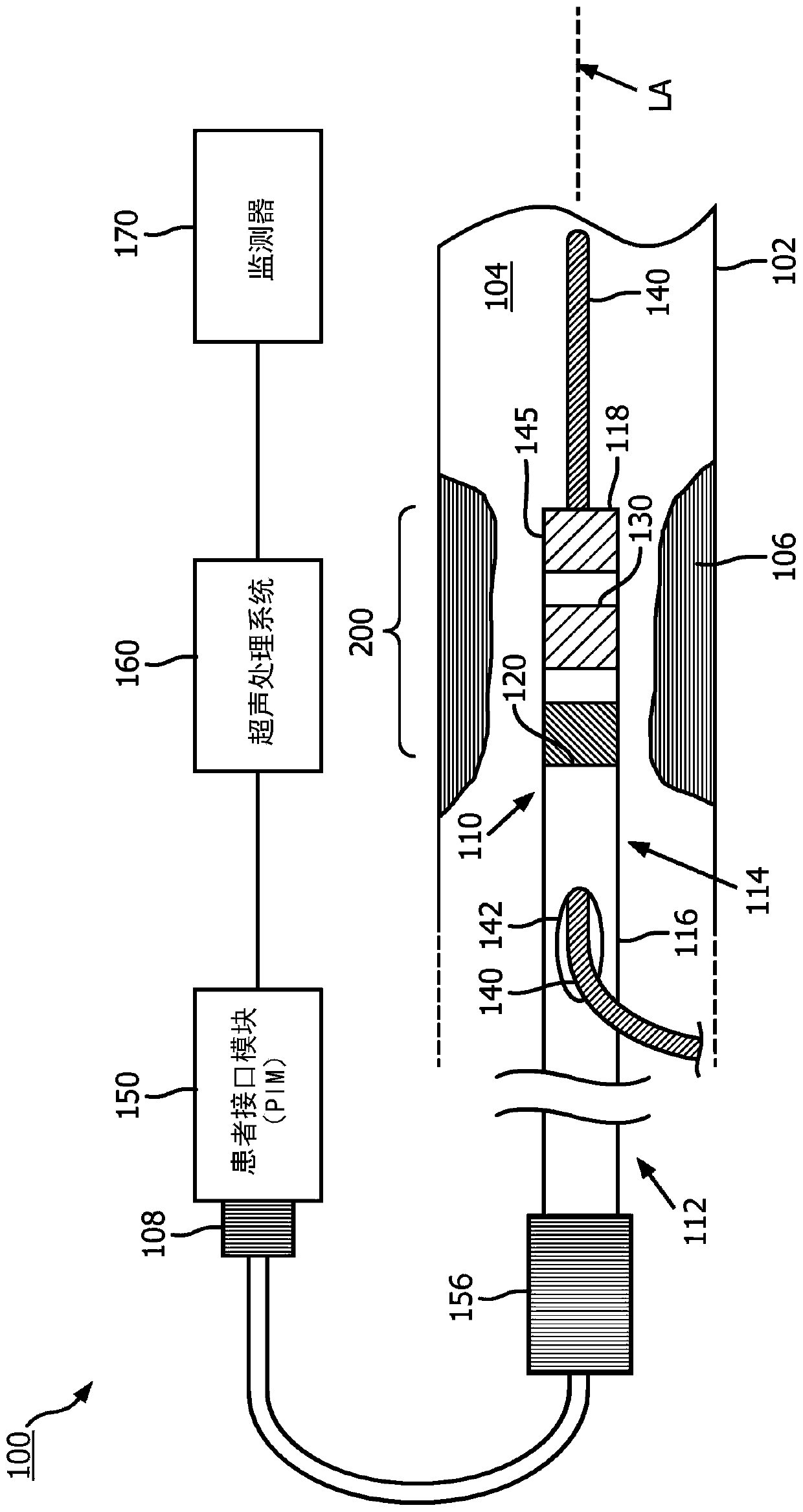
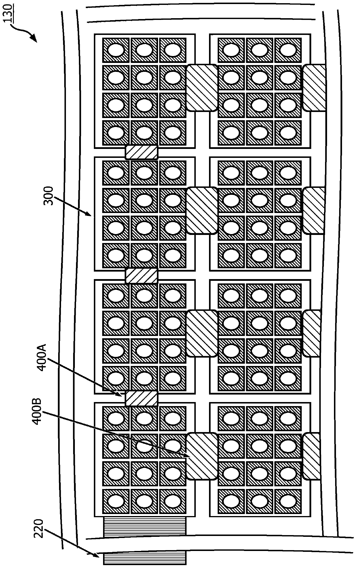
[0017] For the purposes of promoting an understanding of the principles of the disclosure, reference will now be made to the embodiments illustrated in the drawings and specific language will be used to describe the same. It should be understood, however, that no limitation of the scope of the present disclosure is intended. Any alterations and further modifications of the described devices, systems, and methods, and any further applications of the principles of the disclosure are fully contemplated and encompassed within this disclosure, as would normally occur to a person skilled in the art to which this disclosure pertains. For example, while the ICE system is described in terms of intraluminal imaging, it should be understood that it is not intended to limit the application. In particular, it is fully contemplated that features, components and / or steps described with respect to one embodiment may be combined with features, components and / or steps described with respect to ...


PUM


Abstract

Intraluminal ultrasound devices, systems and methods are provided. In one embodiment, an intraluminal ultrasound device includes a flexible elongate member configured to be positioned within a body lumen of a patient, the flexible elongate member including a distal portion and a longitudinal axis; and a transducer array disposed at the distal portion of the flexible elongate member and circumferentially positioned around the longitudinal axis of the flexible elongate member. The transducer array includes a plurality of micromachined ultrasound transducers (MUTs). In addition, the transducer array is configured to obtain ultrasound imaging data of the body lumen in response to a first electrical signal, and apply an ultrasound therapy within the body lumen in response to a second electricalsignal.

Description

technical field [0001] The present disclosure relates generally to intravascular ultrasound devices, and in particular to intraluminal ultrasound devices with ultrasound transducer arrays tunable for both imaging and therapeutic applications. Background technique [0002] Intravascular ultrasound (IVUS) imaging is widely used in interventional cardiology as a diagnostic tool for evaluating diseased blood vessels, such as arteries, in the human body to determine the need for treatment, guide intervention and / or assess its effectiveness. An IVUS device including one or more ultrasound transducers is delivered into the blood vessel and directed to the area to be imaged. The transducer emits ultrasound energy with a frequency above 10 MHz to create an image of the vessel of interest. Ultrasound is caused in part by discontinuous reflections from tissue structures (such as the various layers of blood vessel walls), red blood cells, and other features of interest. Echoes from th...

Claims


Application Information

Patent Timeline
Patent Type & Authority Applications(China)
IPC IPC(8): A61B8/12A61B8/00A61N7/02A61M37/00
CPCA61B8/4483A61B8/56A61M37/0092A61N7/02A61B17/22A61B18/14A61B2018/00577A61B2018/00601A61B2218/007A61N2007/0073A61B34/25A61B2090/378A61B17/2202A61B17/320758A61B8/12A61N7/00A61N2007/0052A61N7/022A61N2007/0043A61B1/015A61B8/4488A61B8/5223A61B8/54A61M25/00
Inventor R·E·卡尼J·斯蒂加尔P·萨罗哈
Owner KONINKLJIJKE PHILIPS NV
Why Eureka
Try Eureka