Application of deep learning for medical imaging evaluation

A deep learning, medical imaging technology, applied in medical imaging, understanding medical/anatomical patterns, applications, etc., can solve complex problems

Pending Publication Date: 2020-07-21
库雷人工智能科技私人有限公司
View PDF6 Cites 0 Cited by
  • Summary
  • Abstract
  • Description
  • Claims
  • Application Information

AI Technical Summary

Problems solved by technology

While trials are often performed, reading chest radiographs becomes a more complex radiological task and is notoriously highly subjective, with differences between reviewers depending on their level of experience, abnormalities detected, and clinical setting The agreement varies from a kappa value of 0.2 to a kappa value of 0.77

Method used


Image

  • Application of deep learning for medical imaging evaluation
  • Application of deep learning for medical imaging evaluation
  • Application of deep learning for medical imaging evaluation

Examples


example 1

[0046] Example 1 Radiologist Validation of a Deep Learning System to Detect Chest X-ray Abnormalities
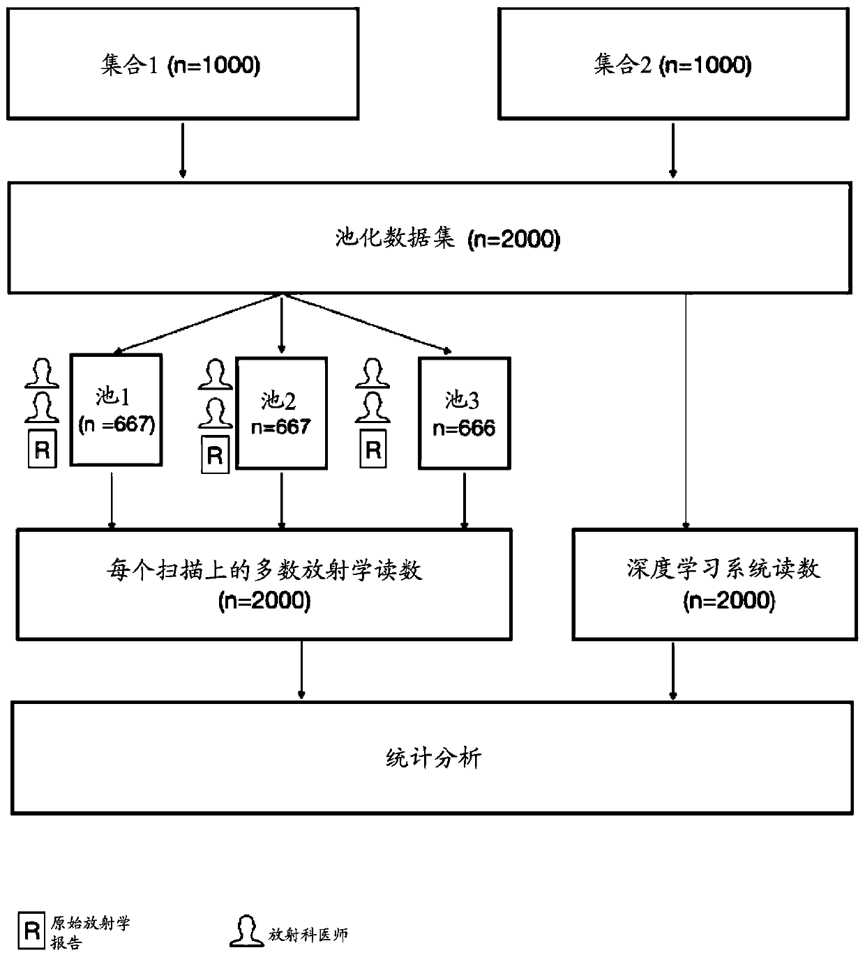
[0047] 1.1. Method

[0048] 1.1.1 Algorithm Development

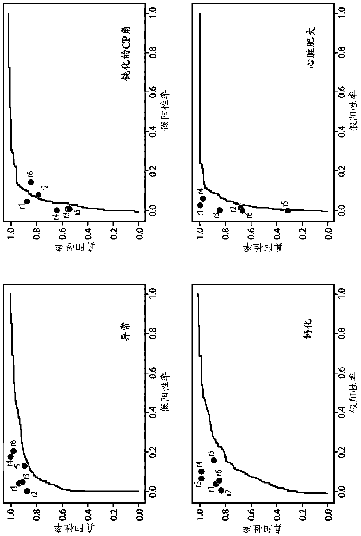
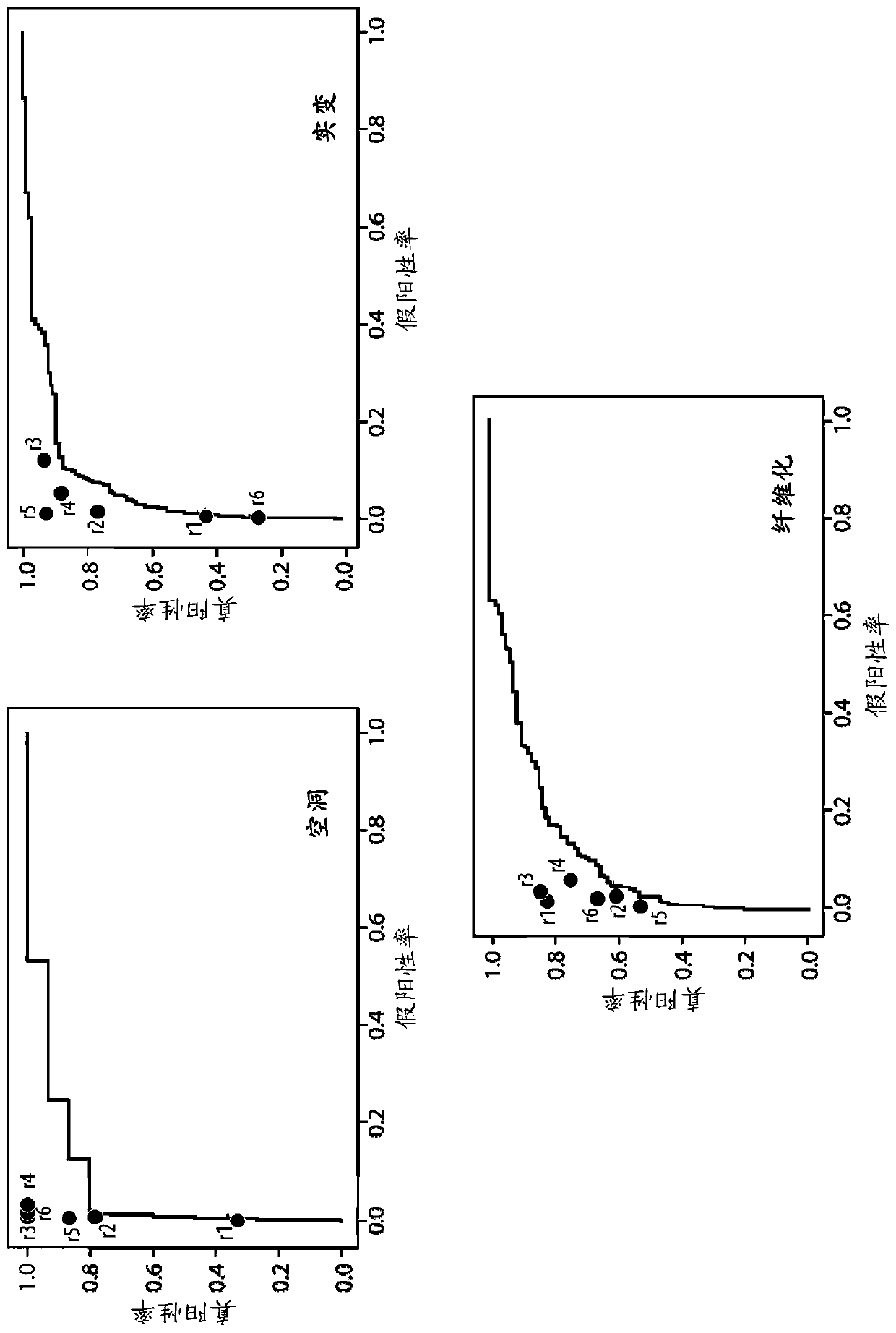
[0049] 1,200,000 radiographs and their corresponding radiology reports were used to train a convolutional neural network (CNN) to identify abnormalities. A natural language processing algorithm was developed to parse unstructured radiology reports and extract information about the presence of abnormalities in chest radiographs. These extracted findings are used as labels when training the CNN. A single network was trained to recognize normal radiographs, as well as chest radiograph findings "blunted CP angle", "calcification", "cardiac hypertrophy", "cavitation", "consolidation", "fibrosis", " enlarged hilum", "cloudiness" and "pleural effusion". Table 1 lists the definitions used when extracting radiological findings from the reports. These findings are called tags. Label extraction accuracy is measured relativ...

example 2

[0109] Example 2 Deep Learning Solution qXR for Tuberculosis Detection

[0110] Qure.ai's qXR is designed to screen and prioritize abnormal chest X-rays. Algorithms automatically identify the 15 most common chest X-ray abnormalities. A subset of these anomalies suggestive of typical or atypical tuberculosis were grouped together to generate a "TB Screening" algorithm within the results. The TB screening algorithm is designed to replicate the screening of radiologists or physicians' chest radiographs for abnormalities suggestive of TB prior to microbial confirmation. qXR is the first CE-certified AI-based chest X-ray interpretation software. qXR is integrated with the Vendor Neutral Integration Process and works with radiographs generated from any radiograph system (CR or DR). qXR screens for TB and also identifies 15 other abnormalities, so patients can be informed of the non-TB condition they may have.

[0111] qXR integrates seamlessly with Vendor Neutral Archives (VNA) ...


PUM


Abstract

This disclosure generally pertains to methods and systems for processing electronic data obtained from imaging or other diagnostic and evaluative medical procedures. Certain embodiments relate to methods for the development of deep learning algorithms that perform machine recognition of specific features and conditions in imaging and other medical data. Another embodiment provides systems configured to detect and localize medical abnormalities on medical imaging scans by a deep learning algorithm.

Description

[0001] related application [0002] This application claims the benefit of priority from Indian Patent Application No. 201821042893 filed on November 14, 2018, which is hereby incorporated by reference in its entirety for all purposes. technical field [0003] The present disclosure generally relates to methods and systems for processing electronic data obtained from imaging or other diagnostic and evaluation medical procedures. Some embodiments relate to methods for the development of deep learning algorithms that perform machine recognition of specific features and conditions in imaging and other medical data. Background technique [0004] Medical imaging techniques such as computed tomography (CT) and X-ray imaging are widely used in diagnosis, clinical research and treatment planning. There is an emerging need for automated methods that improve the efficiency, accuracy, and cost-effectiveness of medical imaging assessments. [0005] Chest X-rays are among the most comm...

Claims


Application Information

Patent Timeline
Patent Type & Authority Applications(China)
IPC IPC(8): G06K9/00A61B5/05G06V10/82G06V30/12
CPCA61B5/7267A61B2576/00G06V2201/03G06V30/12G06V10/82
Inventor 普雷塔姆·普萨马努基·塔德帕里巴尔加瓦·雷迪塔伦·尼姆马达普贾·拉奥普拉桑特·瓦瑞尔
Owner 库雷人工智能科技私人有限公司
Why Eureka
Try Eureka