
Image region division method and device, equipment and storage medium

An image area and storage medium technology, applied in image analysis, image enhancement, image data processing, etc., can solve problems such as error, long time, and inability to quickly divide the lung lobe area

Pending Publication Date: 2020-09-04
WEBANK (CHINA)
View PDF1 Cites 0 Cited by
  • Summary
  • Abstract
  • Description
  • Claims
  • Application Information

AI Technical Summary

Problems solved by technology

However, in any case, there are certain errors in the automatic segmentation algorithm, and the current segmentation method takes too long to quickly divide the accurate lung lobe area
Especially for patients with adhesions in the lungs or severe emphysema, the lung image segmentation method can no longer be used to segment lung images, which cannot meet the needs of clinical quantitative analysis of diseases

Method used


Image

  • Image region division method and device, equipment and storage medium
  • Image region division method and device, equipment and storage medium
  • Image region division method and device, equipment and storage medium

Examples


Embodiment Construction

[0022] It should be understood that the specific embodiments described herein are only used to explain the present invention, but not to limit the present invention.

[0023] It should be noted that the image area dividing device in the embodiment of the present invention may be devices such as a smart phone, a personal computer, and a server, and there is no specific limitation here.

[0024] The execution subject of the image area division method provided by the embodiment of the present invention may be any image processing device. For example, the image area division method may be executed by an image area division device or a server, where the image area division device may be a user equipment (User Equipment). Equipment, UE), mobile devices, user terminals, terminals, cellular phones, cordless phones, personal digital assistants (PDAs), handheld devices, computing devices, vehicle-mounted devices, wearable devices, etc. The server can be a local server or a cloud server. In ...


PUM


Abstract

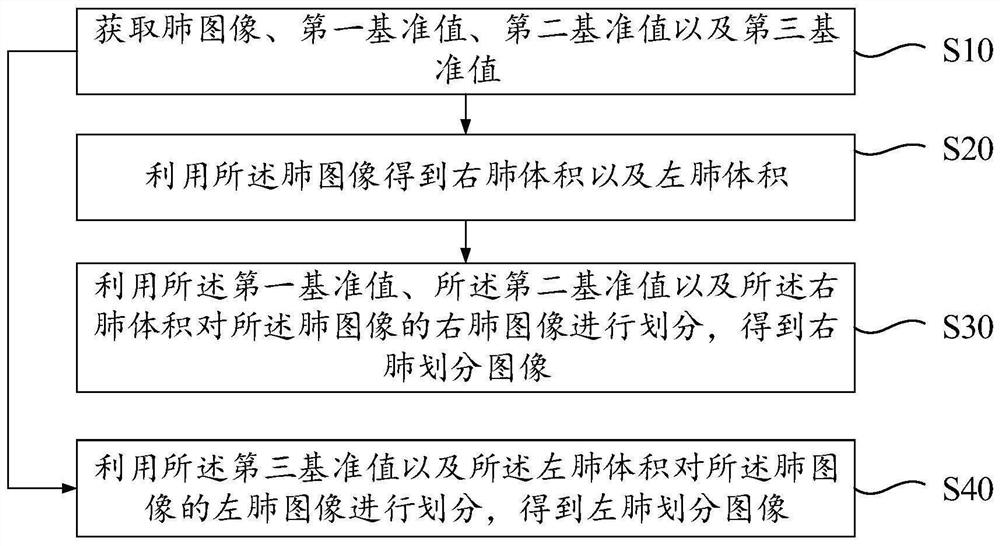
The invention discloses an image region division method, apparatus and device, and a storage medium. The method comprises the steps of obtaining a lung image, a first reference value, a second reference value and a third reference value; utilizing the lung image to obtain a right lung volume and a left lung volume; dividing a right lung image of the lung image by using the first reference value, the second reference value and the right lung volume to obtain a right lung division image; and dividing a left lung image of the lung image by using the third reference value and the left lung volumeto obtain a left lung division image. The lung lobe area can be rapidly and accurately divided, and rapid quantitative analysis of clinical diseases can be achieved in an assisted mode.

Description

Technical field [0001] The present invention relates to the field of artificial intelligence technology, in particular to an image area division method, device, equipment and storage medium. Background technique [0002] The division of lobes of the lung is an important topic in the respiratory field. The purpose of division of the lobes of the lung is to determine the specific distribution of a certain disease in a certain lobe for quantitative analysis of the disease. Currently, the lung lobe area is divided by mature automatic segmentation algorithms, such as the watershed algorithm. However, in any case, the automatic segmentation algorithm has certain errors, and the current segmentation method takes too long to quickly divide the accurate lung lobe area. Especially for patients with lung adhesions or severe emphysema, lung lobes segmentation methods can no longer be used to segment lung images, which cannot meet the needs of clinical quantitative analysis of diseases. The...

Claims


Application Information

Patent Timeline
IPC IPC(8): G06T7/11G06T7/62G06T7/00G06T5/00G06T5/50G06N3/04G06N3/08
CPCG06T7/11G06T7/62G06T7/0012G06T5/50G06N3/08G06T2207/10081G06T2207/20081G06T2207/20221G06T2207/30061G06N3/045G06T5/80
Inventor 蔡杭李月
Owner WEBANK (CHINA)
Learn More