Pathology predictions on unstained tissue

A technology for stains, tissue samples, applied in the field of digital pathology, which can solve the problems of inaccurate real labels and masks

Active Publication Date: 2020-09-29
VERILY LIFE SCI LLC
View PDF9 Cites 2 Cited by
  • Summary
  • Abstract
  • Description
  • Claims
  • Application Information

AI Technical Summary

Problems solved by technology

However, morphological differences in serial slices are still significant and may lead to inaccuracies in the ground truth labels and masks generated in this way

Method used


Image

  • Pathology predictions on unstained tissue
  • Pathology predictions on unstained tissue
  • Pathology predictions on unstained tissue

Examples


Embodiment Construction

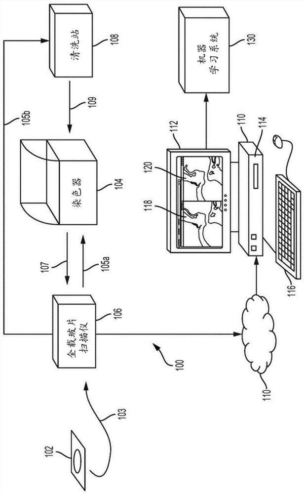
[0032] now turn your attention to figure 1 , figure 1 is an illustration of a laboratory 100 environment in which methods as described herein are practiced. A tissue sample (e.g., a sample that has been formalin-fixed and paraffin-embedded) is placed on a microscope slide 102 and the tissue sample is positioned as shown at 103 to be presented to a whole slide scanner 106 . Such scanners are also well known and available from various suppliers. The whole slide scanner 106 scans the slide at a user-specified magnification, such as 10X, 20X, or 40X. Whole slide scanners include a digital camera for capturing magnified color digital images of the specimen. A digitally enlarged image of the unstained slide (“unstained image”) is then stored locally in the whole slide scanner 106, or on the local hard drive 114 of the pathology workstation 110 in a cloud network or other remote server , or some other storage medium.

[0033] After the slide is scanned by the whole slide scan...


PUM


Abstract

A method for training a pattern recognizer to identify regions of interest in unstained images of tissue samples is provided. Pairs of images of tissue samples are obtained, each pair including an unstained image of a given tissue sample and a stained image of the given tissue sample. An annotation (e.g., drawing operation) is then performed on the stained image to indicate a region of interest. The annotation information, in the form of a mask surrounding the region of interest, is then applied to the corresponding unstained image. The unstained image and mask are then supplied to train a pattern recognizer. The trained pattern recognizer can then be used to identify regions of interest within novel unstained images.

Description

[0001] Cross References to Related Applications [0002] This application claims priority to U.S. Provisional Patent Application No. 62 / 631,259, filed February 15, 2018, which is hereby incorporated by reference. Background technique [0003] The present disclosure relates to the field of digital pathology, and more particularly, to a method for generating a mask in a digital image of a tissue specimen. The term "mask" in this document refers to a closed polygonal region or other specified region in an image of a tissue specimen that encloses or otherwise indicates a region of interest, such as cancer cells (eg, tumor cells). [0004] Digital images of tissue samples with masks and possibly associated labels of the samples (such as "cancerous") are used in several contexts, including as training examples for constructing machine learning models. Such machine learning models can be developed for a variety of purposes, including aiding in diagnosis, clinical decision support, a...

Claims


Application Information

Patent Timeline
Patent Type & Authority Applications(China)
IPC IPC(8): G06K9/00G01N1/30G06K9/32G06T7/00G06V10/25
CPCG01N1/30G06T7/0012G06V20/695G06V10/25G06T2207/30024G06T2207/20081G06T2207/20084G06T2207/30068
Inventor M.斯坦普L.彭
Owner VERILY LIFE SCI LLC
Why Eureka
Try Eureka