
System and method for accelerated clinical workflow

A subsystem and imaging system technology, applied in image analysis, image enhancement, instruments, etc., can solve a large number of computing tasks and other problems

Pending Publication Date: 2020-10-20
GENERAL ELECTRIC CO
View PDF0 Cites 0 Cited by
  • Summary
  • Abstract
  • Description
  • Claims
  • Application Information

AI Technical Summary

Problems solved by technology

Furthermore, in many cases quantization is computationally intensive and may need to be performed offline

Method used


Image

  • System and method for accelerated clinical workflow
  • System and method for accelerated clinical workflow
  • System and method for accelerated clinical workflow

Examples


Embodiment Construction

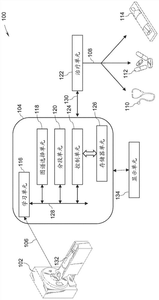
[0013] As will be described in detail below, systems and methods for accelerating clinical workflow are presented. More specifically, systems and methods for accelerating radiology workflow using deep learning are presented.

[0014] It should be understood that radiology is the science of using X-ray imaging to examine and record medical conditions in a subject, such as a patient. Some non-limiting examples of medical techniques used in radiology include radiography, ultrasound, computed tomography (CT), positron emission tomography (PET), magnetic resonance imaging (MRI), and the like. The term "anatomical image" refers to a two-dimensional image representing anatomical regions within a patient such as, but not limited to, the heart, brain, kidneys, prostate, and lungs. Furthermore, the terms "segmented image", "segmented image" and "sub-segmented image" are used herein to refer to anatomical images in which parts and / or sub-regions of anatomical structures are border-label...


PUM


Abstract

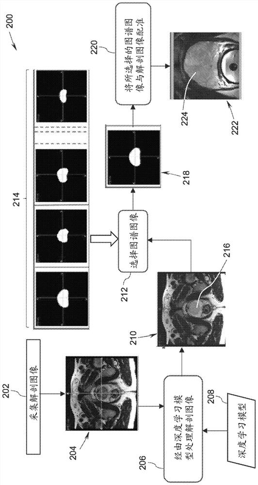
A method for imaging configured to provide an accelerated clinical workflow includes acquiring anatomical image data corresponding to an anatomical region of interest in a subject. The method furtherincludes determining localization information corresponding to the anatomical region of interest based on the anatomical image data using a learning based technique. The method also includes selectingan atlas image from a plurality of atlas images corresponding to the anatomical region of interest based on the localization information. The method further includes generating a parcellated segmented image based on the atlas image. The method also includes recommending a medical activity based on the parcellated segmented image. The medical activity includes at least one of guiding image acquisition, supervising a treatment plan, assisting therapeutic delivery to the subject, and generating a medical report.

Description

Background technique [0001] Embodiments of the present specification relate generally to clinical workflow, and more specifically to systems and methods for accelerating radiology workflow using deep learning. [0002] Medical imaging data are increasingly used in the diagnosis and treatment of health conditions, such as, but not limited to, cancer conditions and arterial disease. Imaging techniques such as computed tomography (CT) and magnetic resonance imaging (MRI) generate a large number of medical images with valuable diagnostic information. Acquiring medical image data associated with anatomical regions within a patient may require repeated imaging. Additionally, these images need to be analyzed by a medical professional to derive any usable diagnostic information. Additionally, the study of images is a laborious and time-consuming process. [0003] Automatic segmentation and analysis of medical image volumes is a promising and valuable tool for medical professionals ...

Claims


Application Information

Patent Timeline
IPC IPC(8): G06T7/11G06T7/174
CPCG06T7/11G06T7/174G06T2207/20081G06T2207/20128G06T2207/30081G06T2207/20084
Inventor 达蒂什·达亚南·尚巴格拉凯什·穆利克克里希纳·希瑟拉姆·施莱姆桑迪普·苏亚纳拉亚纳·考希克阿拉蒂·斯雷库马里
Owner GENERAL ELECTRIC CO
Learn More