
Automated qualitative description of anatomical changes in radiotherapy

a radiotherapy and qualitative technology, applied in the field of automatic monitoring of anatomical changes, can solve the problems of time-consuming, manual processing and interpretation, visual assessment, and affecting the anatomy of the patient, and achieve the effect of convenient for the physician and faster and more accurate delivery

Pending Publication Date: 2022-02-24
KONINKLJIJKE PHILIPS NV
View PDF0 Cites 0 Cited by
  • Summary
  • Abstract
  • Description
  • Claims
  • Application Information

AI Technical Summary

Benefits of technology

The system described in this patent allows for faster and more accurate monitoring of anatomical changes in patients undergoing radiation therapy. By using a qualitative scoring system, changes are identified and described in a way that is easy to understand and can be further analyzed. This information, presented in a user-friendly way, can be used to make better treatment decisions. The system can also help with consistent and accurate monitoring over time, which can improve patient outcomes.

Problems solved by technology

Each fraction of the therapy that is delivered to a patient can also impact the anatomy.
However, visual assessment, manual processing and interpretation are error prone and time consuming.
Errors can occur, because the clinician may overlook anatomical changes that are relevant, or can erroneously interpret the spatial relationships between objects in different images.
Also, manual entry of qualitative descriptions can lead to variations in description of similar situations, which can introduce inconsistencies.
In addition, interpretation of anatomical changes requires the analysis of large amounts of data and specialized skills.

Method used


Image

  • Automated qualitative description of anatomical changes in radiotherapy
  • Automated qualitative description of anatomical changes in radiotherapy
  • Automated qualitative description of anatomical changes in radiotherapy

Examples


Embodiment Construction

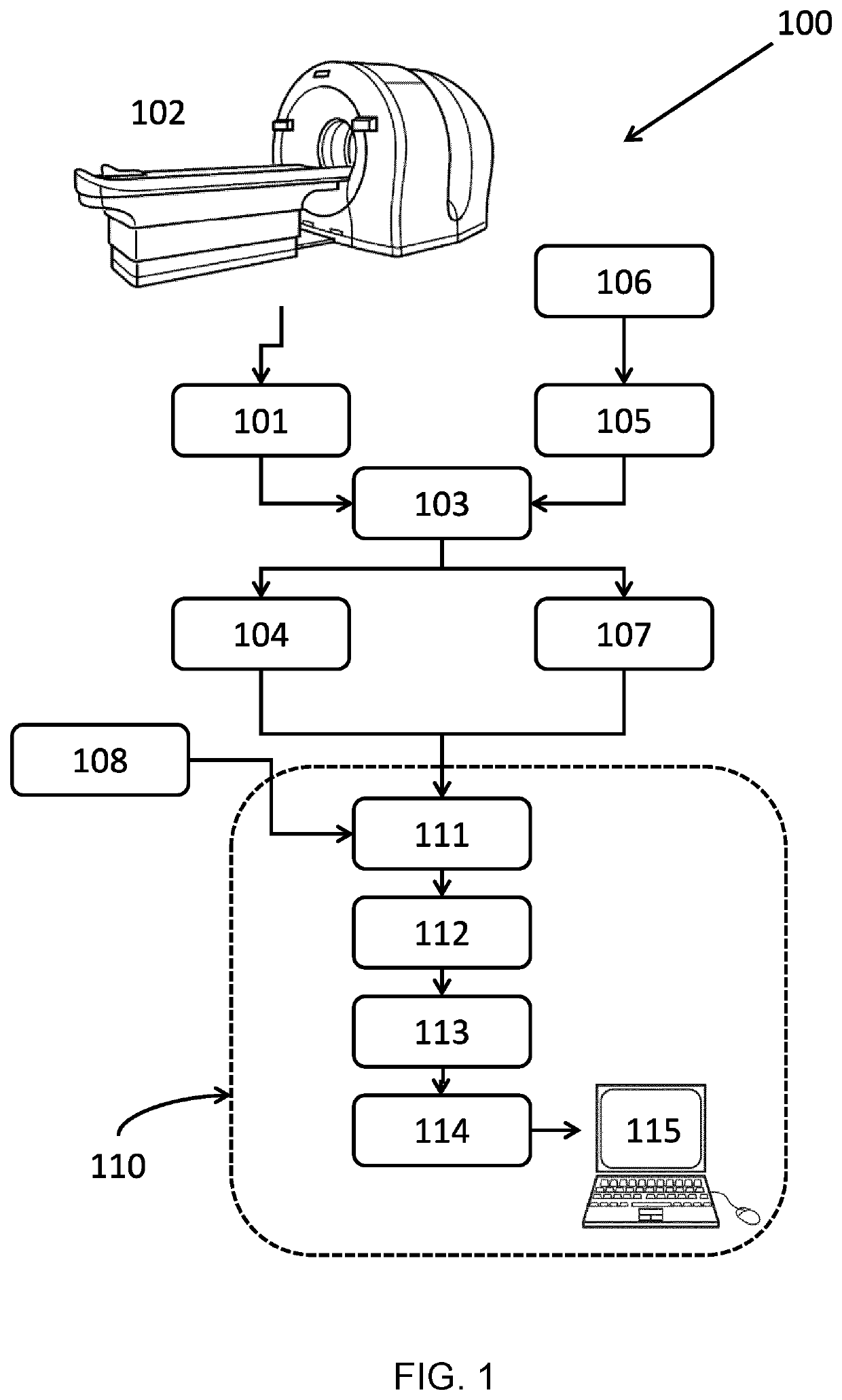
[0030]FIG. 1 illustrates a system for monitoring anatomical changes 110 in a subject in radiation therapy. In this example, the system is illustrated as part of an arrangement 100 for medical imaging and analysis.

[0031]In the arrangement for medical imaging and analysis 100, a planning image 101 is acquired of the subject to be treated by an imaging device 102. The image can be a computed tomography image (CT), magnetic resonance image (MR), positron emission tomography (PET) image, another medical image, or a combined image, such as a combined PET / CT or PET / MR image. In FIG. 1, as an example, a PET / CT imaging device 102 is illustrated. The regions of interest (ROIs) are delineated in this first image using a contouring tool 103 to provide first anatomical image data 104. The ROIs in the subject will comprise at least one target structure (TS), normally a tumor, but may also include one or more organs at risk (OARs). A contouring tool commonly interacts with the user, who can be for...


PUM


Abstract

A system and a method for monitoring anatomical changes in a subject in radiation therapy are provided, as well as an arrangement for medical imaging and analysis and a computer program product for carrying out the method. For monitoring anatomical changes in a subject in radiation therapy, the following steps are performed. First anatomical image data and subsequent anatomical image data of the subject are received. The first anatomical image data and the subsequent anatomical image data are analyzed. This analysis comprises registering the subsequent anatomical data to the first anatomical data. Changes between the first anatomical image data and the subsequent anatomical image data are identified as change states, and the identified change states are matched to corresponding qualitative descriptions. A monitoring report is provided, which comprises the qualitative descriptions of the identified changes.

Description

FIELD OF THE INVENTION[0001]The invention generally relates to the monitoring of anatomical changes, and in particular, but not exclusively, the invention relates to automated monitoring of anatomical changes in subjects undergoing radiation therapy.BACKGROUND OF THE INVENTION[0002]In radiation therapy, target structures in patient's bodies, such as tumors, are treated by subjecting them to radiation. The radiation can be in the form of external radiation such as photons, or particles such as protons, for example in external beam radiotherapy (EBRT). The treatment is delivered in such a way that the radiation that is delivered to the target structures (TSs) is as high as possible, while at the same time the radiation delivered to the surrounding healthy tissue and structures, usually referred to as organs at risk (OARs), is as low as possible.[0003]During the therapy, the dose of radiation is usually delivered to the patient over several radiation therapy sessions, with a recovery p...

Claims


Application Information

Patent Timeline
IPC IPC(8): A61N5/10
CPCA61N5/1049A61N2005/1074
Inventor BONDAR, MARIA LUIZABAL, MATTHIEU FRÉDÉRICISOLA, ALFONSO AGATINO
Owner KONINKLJIJKE PHILIPS NV
Learn More