Laryngoscope lens assembly and tongue depressor

A technology of lens components and spatulas, applied in laryngoscopes, endoscopes, bronchoscopes, etc., can solve the problems of increased virus infection, high consumption of raw materials, and environmental protection, and achieve reduced storage volume, reduced infection rate, avoid cumbersome effects

Pending Publication Date: 2020-10-30
ZHEJIANG UE MEDICAL
View PDF6 Cites 0 Cited by
  • Summary
  • Abstract
  • Description
  • Claims
  • Application Information

AI Technical Summary

Problems solved by technology

To sum up, the traditional tongue depressor has the following disadvantages: there is no direct protective measure between medical staff and patients, which increases the chance of virus infection; when virus collection or pharyngeal examination is performed, an external light source is required due to the influence of the environment, which is not convenient for medical care Personnel operation; wooden tongue depressor consumes a lot of raw materials and is not environmentally friendly

Method used


Image

  • Laryngoscope lens assembly and tongue depressor
  • Laryngoscope lens assembly and tongue depressor
  • Laryngoscope lens assembly and tongue depressor

Examples


Embodiment 1

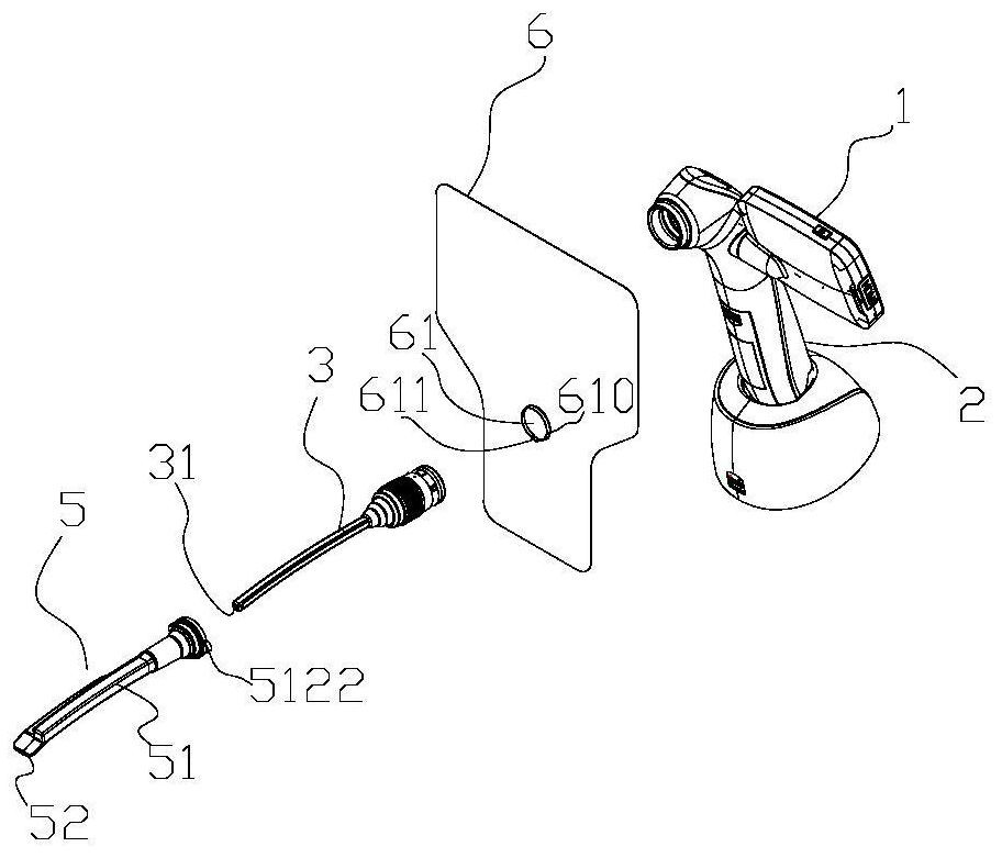
[0025] see Figure 1~2 , the video laryngoscope includes a display 1, a handle 2, a laryngoscope handle 3 and a lens 5, wherein the display 1 and the laryngoscope handle 3 are arranged on the handle 2, and the front end of the laryngoscope handle 3 is equipped with a visual device such as a camera 31, the lens 5 is sleeved on the handle 5 of the laryngoscope in a detachable manner;

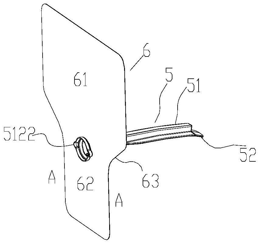
[0026] Among them, refer to Figure 4 The lens 5 includes a tube body 51 for being sleeved on the handle of the laryngoscope and a tongue depressor 52 that is arranged on the tube body 51 and protrudes forward relative to the front end of the tube body 51. The body 51 includes an arc-shaped pipe section 511 adapted to the physiological curvature of the oral cavity and throat of the human body and a connecting pipe section 512 for connecting the handle 3 of the laryngoscope. Detachable connection and coordination;

[0027] Based on the video laryngoscope above, see again Figure 1~2 , the prese...

Embodiment 2

[0044] This embodiment provides a video laryngoscope, including a display 1, a handle 2, a laryngoscope handle 3 and the lens assembly described in Embodiment 1, wherein the display is arranged on the handle for observation by a doctor, and the laryngoscope handle 3 It is arranged at the bottom of the handle and the front end of the handle of the laryngoscope is equipped with a visual device such as a camera, which is used to transmit the image in the patient's body to the display, and the lens 5 is detachably sleeved on the laryngoscope on the mirror handle.

[0045] The lenses and baffles are preferably disposable products.

Embodiment 3

[0047] According to the idea of ​​Example 1, this embodiment provides a spatula, the spatula includes a spatula body, and the side of the spatula body close to the doctor is provided to block the gap between the doctor and the patient. Protection against virus transmission.

[0048] The tongue depressor provided by this embodiment can effectively block the virus transmission between doctors and patients; especially when the doctor collects throat swabs for detection, the spatula provided by the present invention can effectively block other gases and liquids such as aerosols exhaled by patients Transmission to doctors significantly reduces the infection rate of doctors in the diagnosis and treatment of viral infectious diseases; at the same time, it also avoids the transmission of doctors to patients.

[0049] It can be understood that the spatula body can be integrally connected with other structures. As described in Embodiment 1, the spatula and the laryngoscope lens can also...


PUM


Abstract

The invention provides a laryngoscope lens assembly. The laryngoscope lens assembly comprises a lens and a protection piece which is arranged at the rear end of the laryngoscope lens and used for blocking virus transmission between a doctor and a patient.

Description

technical field [0001] The invention belongs to the field of visual medical instruments, and in particular relates to a laryngoscope lens assembly and a tongue depressor. Background technique [0002] The tongue depressor is a device used to depress the tongue to facilitate the inspection of surrounding organs and tissues. It is mainly used for pharyngeal inspection and is an essential inspection tool for doctors. The traditional tongue depressor is a long wooden thin wood chip with semicircular ends. Wooden spatula is a consumable material, which requires a lot of wood. The doctor gently presses the front 2 / 3 of the patient's tongue with a spatula, and checks the tonsils, lingual and palatine arches, pharyngeal and palatine arches, and the posterior pharyngeal wall. During the outbreak, throat swab collection is an important test to detect whether you are infected with the new coronavirus. Collecting throat swabs for nucleic acid testing is one of the methods for diagnosi...

Claims


Application Information

Patent Timeline
Patent Type & Authority Applications(China)
IPC IPC(8): A61B1/267A61B1/00
CPCA61B1/267A61B1/00142A61B1/00163A61B1/00096
Inventor 陈肖敏翁勤张骏王卫东张家智黄运东张玄玄郑志平黄秋媛季颖波吴志坚
Owner ZHEJIANG UE MEDICAL
Why Eureka
Try Eureka