CT image metal artifact removing method and device and computer readable storage medium

A metal artifact, CT image technology, applied in the field of medical imaging, can solve the problem of inability to integrate data integrity information, and achieve the effect of improving accuracy

Active Publication Date: 2020-12-01
NANJING ANKE MEDICAL TECH CO LTD
View PDF4 Cites 6 Cited by
  • Summary
  • Abstract
  • Description
  • Claims
  • Application Information

AI Technical Summary

Problems solved by technology

Secondly, the existing technology in the image domain cannot integrate the data integrity information in the metal scanning data into the process of image domain processing. The integrity of this data is also of great significance to the processing of metal artifacts.

Method used


Image

  • CT image metal artifact removing method and device and computer readable storage medium
  • CT image metal artifact removing method and device and computer readable storage medium
  • CT image metal artifact removing method and device and computer readable storage medium

Examples


Embodiment Construction

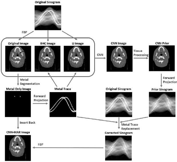
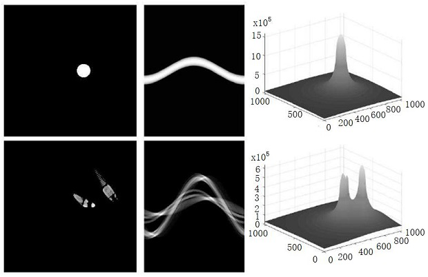
[0042] The present invention proposes a deep learning metal artifact suppression method based on human tissue structure and data integrity, which is used to improve the quality of CT images. The schematic diagram of the metal artifact removal method of the present invention is as follows Figure 4 to Figure 6shown. The purpose of the present invention is to correct metal artifacts by means of deep learning to help doctors diagnose quickly and accurately. Specific steps are as follows:

[0043] 1. Create a data set: including images with metal artifacts and images without metal artifacts, and their corresponding projection images, reference 1. The specific method is to collect different CT images of patients, which are divided into two parts containing metal and not containing metal; segment and extract the metal part from the CT image containing metal implants as a simulated implant; Image processing containing metal artifacts to obtain metal-free projection data; insert si...


PUM


Abstract

The invention discloses a CT image metal artifact removing method and device and a computer readable storage medium, and the method comprises the steps: collecting a metal artifact-containing image and a metal artifact-free image, and processing the metal artifact-containing image and the metal artifact-free image to obtain corresponding projection data; reconstructing the generated two projectiondata respectively to generate an image containing metal artifacts and an image without artifacts, and further processing to obtain an initial artifact suppression image and a data integrity map; anddesigning a neural network and a loss function, wherein the neural network outputs an image after artifact removal and the like. The CT image metal artifact removing method is characterized in that correlation between metal artifacts and a surrounding structure and the relation between metal artifacts and tissue components are utilized, the problem that data integrity information in metal scanningdata cannot be fused into the image domain processing process in the existing image domain technology is solved, and the artifact correction accuracy and the CT image quality are improved.

Description

technical field [0001] The invention relates to the technical field of medical imaging, in particular to a method, a device and a computer-readable storage medium for removing metal artifacts in CT images. Background technique [0002] Computed tomography (CT) can non-destructively detect the tissue and organ structures inside the human body, so it is widely used in clinical diagnosis. Affected by metal implants, different degrees of metal artifacts will appear in CT images, manifested as strip artifacts and bright and dark bands, which seriously affect the image quality and hinder accurate diagnosis by doctors. Therefore, a large number of metal artifacts reduction (MAR) methods have appeared in the past 40 years to remove metal artifacts. Such as data acquisition improvements, physics-based corrections, projection data corrections, iterative methods and image post-processing techniques, and emerging deep learning-based methods. [0003] The improvement method of data acq...

Claims


Application Information

Patent Timeline
Patent Type & Authority Applications(China)
IPC IPC(8): G06T5/00
CPCG06T5/006G06T2207/10081G06T2207/20081G06T2207/20084
Inventor 曾凯冯亚崇
Owner NANJING ANKE MEDICAL TECH CO LTD
Why Eureka
Try Eureka