Method for segmenting capillaries in microscopic image based on generative adversarial network

A microscopic image, generative technology, applied in image analysis, biological neural network model, image enhancement, etc., can solve problems such as low contrast, broken blood vessels, redundant details of blood vessel branches, etc., to remove noise, accelerate training, and enhance The effect of discriminative ability

Inactive Publication Date: 2020-12-11
HARBIN UNIV OF SCI & TECH
View PDF2 Cites 2 Cited by
  • Summary
  • Abstract
  • Description
  • Claims
  • Application Information

AI Technical Summary

Problems solved by technology

[0004] The above method can extract most of the microvascular images. Due to the limitation of the algorithm or the low contrast of the actual imaging

Method used


Image

  • Method for segmenting capillaries in microscopic image based on generative adversarial network
  • Method for segmenting capillaries in microscopic image based on generative adversarial network
  • Method for segmenting capillaries in microscopic image based on generative adversarial network

Examples


specific Embodiment approach 1

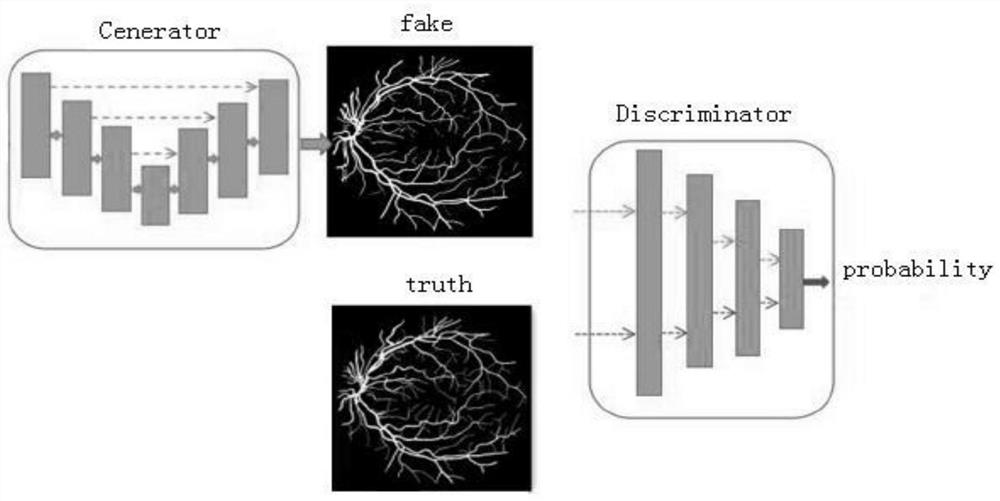
[0040] Specific Embodiments 1. A method for segmenting microvessels in a microscopic image based on a generative confrontation network described in this embodiment, the method includes the following steps:

[0041] Step 1: Establish a training model and sample set based on a generative confrontation network;

[0042] Step 2: Input the color fundus image in the sample set into the generation model, extract the image feature information and output the microvascular probability image under the microscopic image as the generation sample;

[0043] Step 3: Use RGB three-channel microscopic images for network training, and perform enhanced processing on microscopic image contrast adaptive histogram equalization;

[0044] Step 4: Increase the amount of training data to re-enhance the microscopic image processed by the preprocessing unit;

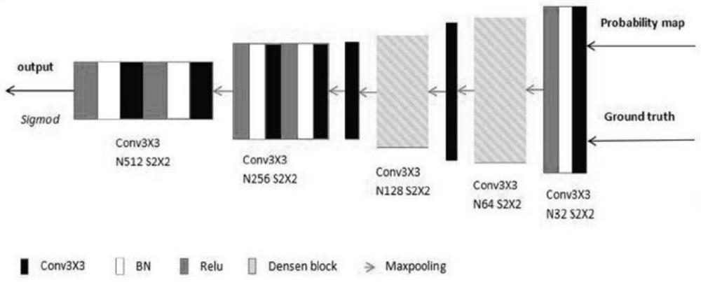
[0045]Step 5: Input the generated sample and the corresponding real sample into the discriminant model at the same time, and the discriminant mode...

specific Embodiment approach 2

[0051] Specific Embodiment 2. This embodiment is a further description of the microvascular segmentation method in microscopic images based on the generative confrontation network described in the specific embodiment 1. The generative network model includes a contraction path, an expansion path, and an output layer.

specific Embodiment approach 3

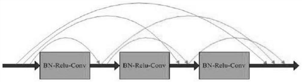
[0052] Specific Embodiment 3. This embodiment is a further description of the microvascular segmentation method in microscopic images based on Generative Adversarial Network described in Embodiment 1. The shrinkage path mainly consists of multiple resnet convolution blocks and Downsampling is composed, and the feature extraction part of the contraction path adopts the idea of ​​resnet network.


PUM


Abstract

The invention discloses a method for segmenting capillaries in a microscopic image based on a generative adversarial network. The objective of the invention is to solve the problems that a vascular fracture phenomenon often occurs in an algorithm segmentation result and vascular branch details are redundant due to the limitation of an algorithm or low actual imaging contrast of a microvascular image. The method comprises the steps of establishing a training model and a sample set based on a generative adversarial network; inputting the color fundus image in the sample set into a generation model, extracting image feature information, and outputting a microvascular probability image under a microscopic image as a generation sample; carrying out enhancement processing on the microscopic image comparison adaptive histogram equalization; increasing the data volume of training, and carrying out enhancement processing on the microscopic image processed by the preprocessing unit again; distinguishing the real sample from the generated sample; inputting a to-be-segmented retinal vessel color image into the segmentation model, and outputting a vessel segmentation result. The method is applied to capillary segmentation in microscopic images.

Description

technical field [0001] The invention relates to a method for segmenting microvessels in microscopic images based on a generative confrontation network. Background technique [0002] Microvascular images under microscopic images have uneven gray distribution, complex vascular structures, low contrast between target blood vessels and image background, and image noise. Microvascular segmentation under microscopic images is facing great challenges. Traditional segmentation methods include methods based on pattern recognition (supervised classification and unsupervised classification), methods based on matched filtering, methods based on mathematical morphology, and methods based on tracking. [0003] Generative Adversarial Network (GAN), the problem to be solved is how to generate new samples that conform to the probability distribution of real samples. An adversarial network can be thought of as consisting of a generative model and a discriminative model. During the training ...

Claims


Application Information

Patent Timeline
IPC IPC(8): G06T7/10G06T5/00G06N3/04G06N3/08
CPCG06T7/10G06T5/007G06N3/084G06T2207/10024G06T2207/10056G06T2207/20081G06T2207/20084G06T2207/30041G06T2207/30101G06N3/045
Inventor 罗中明骆佳楠
Owner HARBIN UNIV OF SCI & TECH
Why Eureka
Try Eureka