Kidney tumor segmentation method based on mixed dimension convolution

A kidney tumor and convolution technology, applied in the field of medical image processing, can solve the problem of increasing the difficulty of feature learning generalization, and achieve the effect of suppressing channels with irrelevant features and good learning effect

Pending Publication Date: 2020-12-15
XIAMEN UNIV
View PDF0 Cites 2 Cited by
  • Summary
  • Abstract
  • Description
  • Claims
  • Application Information

AI Technical Summary

Problems solved by technology

At present, CT imaging examination is one of the main examination methods for renal tumors and other renal diseases. According to the size of renal tumors, doctors can grade the severity of tumors and formulate corresponding treatment methods; at the same time, they can locate renal tumors and analyze their shape and size; existing kidney images obtained through medical image processing are used to accurately segment and judge kidney and kidney tumor regions, which effectively alleviates the workload

Method used


Image

  • Kidney tumor segmentation method based on mixed dimension convolution
  • Kidney tumor segmentation method based on mixed dimension convolution
  • Kidney tumor segmentation method based on mixed dimension convolution

Examples


Embodiment

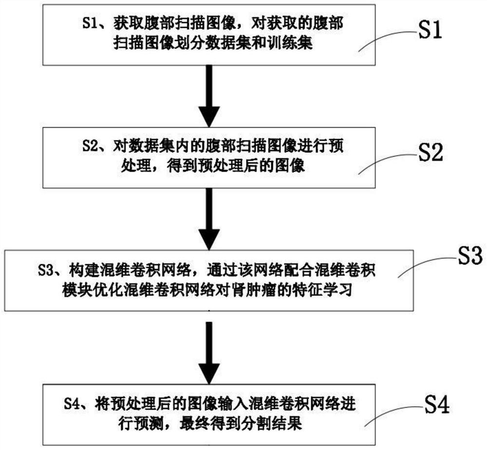
[0037] Cooperate Figure 1 to Figure 5 As shown, the present invention discloses a kidney tumor segmentation method based on mixed-dimensional convolution, comprising the following steps:

[0038] S1. Obtain an abdominal scan image, and divide the acquired abdominal scan image into a data set and a training set.

[0039] S2. Preprocessing the abdominal scan images in the data set to obtain preprocessed images.

[0040] S3. Construct a Mix-dimension Convolution Network (MDC-Net), and use the network to cooperate with a Mix-dimension Convolution block (MDC block) to optimize the feature learning of the mixed-dimension convolution network for renal tumors .

[0041] S4. Input the preprocessed image into the mixed-dimensional convolutional network for prediction, and finally obtain the segmentation result.

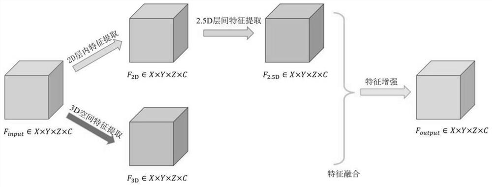
[0042] Cooperate Figure 2 to Figure 3 As shown, the preprocessing in step S2 adopts a downsampling operation, specifically downsampling the acquired abdominal scan image ...


PUM


Abstract

The invention discloses a kidney tumor segmentation method based on mixed dimension convolution, and the method comprises the following steps: S1, obtaining an abdomen scanning image, and dividing theobtained abdomen scanning image into a data set and a training set; S2, preprocessing the abdomen scanning image in the data set to obtain a preprocessed image; S3, constructing a mixed-dimension convolution network, and optimizing the feature learning of the mixed-dimension convolution network for the kidney tumor through the network in cooperation with a mixed-dimension convolution module; S4,inputting the preprocessed image into a mixed-dimension convolutional network for prediction, and finally obtaining a segmentation result; 2D, 2.5 D and 3D convolution features of the kidney tumor arelearned at the same time through the hybrid convolution network, and the generalization ability of model features is enhanced through feature fusion of the 2D, 2.5 D and 3D convolution features.

Description

technical field [0001] The present invention relates to the technical field of medical image processing, in particular to a kidney tumor segmentation method based on mixed-dimensional convolution. Background technique [0002] The kidney is an important organ of the human body. Once the kidney function is damaged, a variety of metabolic end products will accumulate in the body, which will affect life safety. Among various kidney diseases, kidney tumor is the number one dangerous disease for kidney health. At present, CT imaging examination is one of the main examination methods for renal tumors and other renal diseases. According to the size of renal tumors, doctors can grade the severity of tumors and formulate corresponding treatment methods; at the same time, they can locate renal tumors and analyze their shape and size; existing kidney images obtained through medical image processing are used to accurately segment and judge kidney and kidney tumor regions, which effecti...

Claims


Application Information

Patent Timeline
IPC IPC(8): G06T7/10G06T9/00G06N3/04G06N3/08
CPCG06T7/10G06T9/002G06N3/049G06N3/084G06T2207/20081G06T2207/20084G06T2207/30084G06T2207/30096G06T2207/20221G06T2207/10081G06N3/048G06N3/044G06N3/045
Inventor 王连生
Owner XIAMEN UNIV
Why Eureka
Try Eureka