Shear wave elasticity imaging method, ultrasonic imaging system and computer readable storage medium

A technology of elastography and shear wave, which is applied in the medical field, can solve problems such as differences in elastic measurement results, inaccuracy, and unstable tissue elastic measurement values, and achieve the effects of improving stability, convenience and accuracy, and improving accuracy

Pending Publication Date: 2021-02-23
SHENZHEN MINDRAY BIO MEDICAL ELECTRONICS CO LTD
View PDF0 Cites 5 Cited by
  • Summary
  • Abstract
  • Description
  • Claims
  • Application Information

AI Technical Summary

Problems solved by technology

However, in the clinical application of shear wave elasticity, the tissue elasticity measured by the thyroid, breast, muscle and other tissues will become unstable / inaccurate due to the change of tissue elasticity due to the different degrees of tightness of the probe.
How to standardize the pressure range of the probe, there is no similar technology on the market at present, only relying on the doctor's operating experience, so the elastic measurement results obtained by different operators will also have relatively large differences

Method used

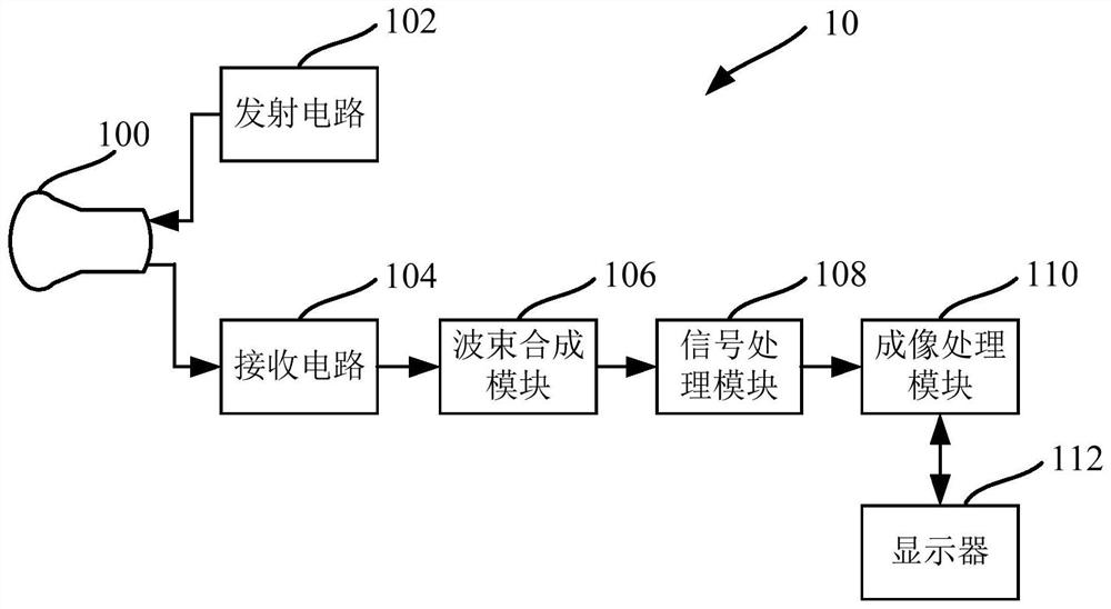

Image

  • Shear wave elasticity imaging method, ultrasonic imaging system and computer readable storage medium
  • Shear wave elasticity imaging method, ultrasonic imaging system and computer readable storage medium
  • Shear wave elasticity imaging method, ultrasonic imaging system and computer readable storage medium

Examples


Embodiment Construction

[0043] In order to enable those skilled in the art to better understand the solution of the present application, the technical solution in the embodiment of the application will be clearly and completely described below in conjunction with the accompanying drawings in the embodiment of the application. Obviously, the described embodiment is only It is a part of the embodiments of this application, not all of them. Based on the embodiments in this application, all other embodiments obtained by persons of ordinary skill in the art without creative efforts fall within the protection scope of this application.

[0044] The terms "first", "second" and the like in the specification and claims of the present application and the above drawings are used to distinguish different objects, rather than to describe a specific order. Furthermore, the terms "comprising" and "having", as well as any variations thereof, are intended to cover a non-exclusive inclusion. For example, a process, m...


PUM


Abstract

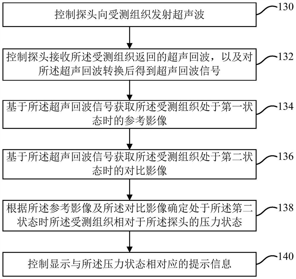
The embodiment of the application discloses a shear wave elasticity imaging method and system and a computer readable storage medium. The method comprises the following steps: controlling a probe to transmit ultrasonic waves to tested tissue; receiving an ultrasonic echo returned by the tested tissue, and controlling the probe to convert the ultrasonic echo to obtain an ultrasonic echo signal; acquiring a reference image of the tested tissue when the tested tissue is in the first state based on the ultrasonic echo signal; acquiring a comparison image of the tested tissue when the tested tissueis in a second state based on the ultrasonic echo signal in a shear wave imaging mode; determining the pressure state of the tested tissue relative to the probe when the tested tissue is in the second state according to the reference image and the comparison image; and controlling display of prompt information corresponding to the pressure state. According to the shear wave elasticity imaging method, the ultrasonic imaging system and the computer readable storage medium, the pressure state between the probe and the tested tissue is determined through the ultrasonic image, and the corresponding prompt information is output, so that a user can conveniently determine the accuracy of the obtained elasticity information of the tested tissue according to the prompt information.

Description

technical field [0001] The present application relates to the medical field, in particular to a shear wave elastic imaging method, an ultrasonic imaging system and a computer-readable storage medium. Background technique [0002] Shear wave elastography generates acoustic radiation force by emitting a focused ultrasound beam, forming a shear wave source in tissue and generating transversely propagating shear waves. By identifying and detecting the shear wave generated inside the tissue and its propagation parameters and imaging these parameters, the difference in tissue stiffness can be quantitatively and visually obtained. Since the excitation of the shear wave comes from the acoustic radiation force generated by the focused ultrasound beam and no longer depends on the pressure applied by the operator, the shear wave elastography method is more stable and reliable than conventional elastography. Reproducibility and other aspects have been improved. However, in the clinica...

Claims


Application Information

Patent Timeline
Patent Type & Authority Applications(China)
IPC IPC(8): A61B8/00
CPCA61B8/485
Inventor 李双双兰帮鑫
Owner SHENZHEN MINDRAY BIO MEDICAL ELECTRONICS CO LTD
Why Eureka
Try Eureka