Surgical vascular endoscope device and working method thereof
A technique for surgery and blood vessels, applied in the field of vascular endoscopy, can solve the problems of unreasonable inflation, high use cost, poor observation effect, etc., and achieve the effects of sufficient light, preventing blockage, and reducing use cost.
- Summary
- Abstract
- Description
- Claims
- Application Information
AI Technical Summary
Problems solved by technology
Method used
Image

Examples
Embodiment 1
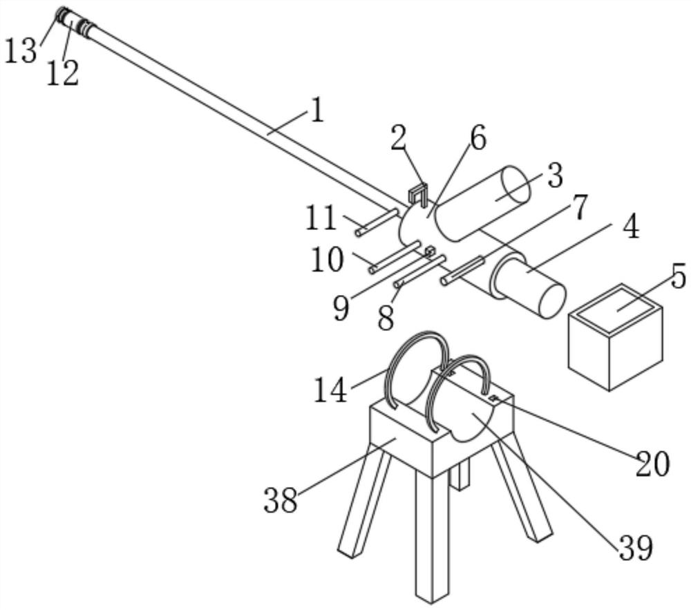
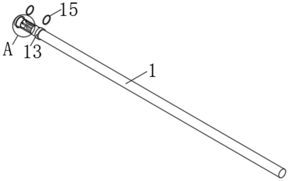
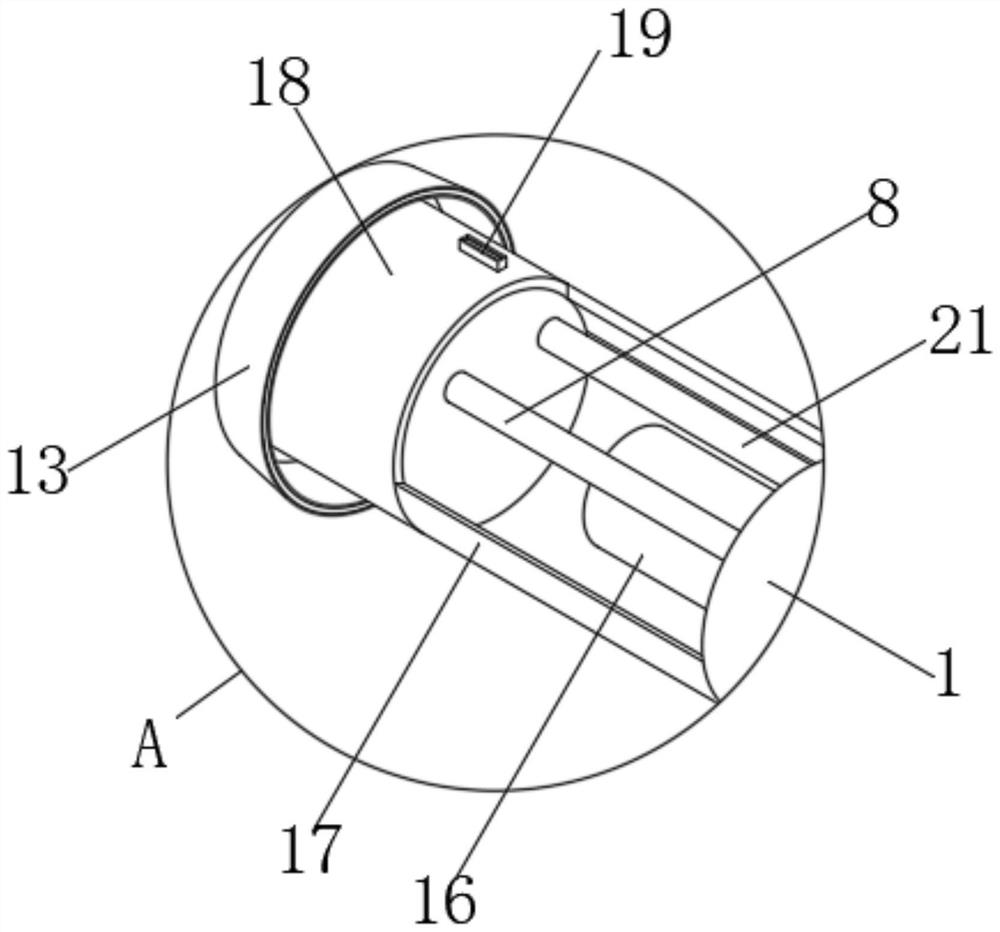
[0041] In a typical implementation of the present application, such as Figure 1-10 As shown, a kind of endoscopic equipment for surgery includes a mirror body 1, the mirror body 1 is a hollow arc-shaped cylindrical structure, two ends of the mirror body 1 are provided with a number of installation holes, the inner wall surface of the mirror body 1 and the outer wall surface A liquid pipeline 28 is opened between them, and a connection part 6 is fixedly installed on the right end of the mirror body 1. The connection part 6 is a cylindrical structure, and the outer wall surface of the connection part 6 is provided with a number of through holes. The handle 3 and the control handle 3 are existing structures, and will not be described in detail here. The outer wall surface of the connecting part 6 is fixedly equipped with a light source inlet 7, and the light source inlet 7 is a cylindrical structure, and the outer wall surface of the connecting part 6 is fixedly installed with a ...
Embodiment 2
[0051] In order to overcome the deficiencies in the prior art, the present invention also provides a working method of a surgical endoscopic device, the specific steps are as follows:
[0052] S1: First insert the mirror body 1 into the blood vessel to be operated, inject gas into the gas injection tube 10, and then the gas enters the transparent air bag 12 from the gas outlet 32 along the pipe, and the transparent air bag 12 is squeezed by the gas and starts to expand. The distance monitor 25 on the inner wall of the transparent airbag 12 will monitor the distance between the inner walls of the transparent airbag 12 at all times. When the distance between the inner walls of the transparent airbag 12 does not change much, it means that the transparent airbag 12 has expanded to the extent that it fits the blood vessel , now the transparent air bag 12 will transmit the signal to the alarm 9, and the alarm 9 makes a sound, and now the gas injection can be stopped.
[0053] S2: ...
PUM
 Abstract
Description
Claims
Application Information