Method for segmenting and marking multi-branch tubular structure in three-dimensional image

A tubular structure, three-dimensional image technology, applied in the field of medical image processing, can solve problems such as blank space

Active Publication Date: 2021-03-05
TSINGHUA UNIV
View PDF3 Cites 6 Cited by
  • Summary
  • Abstract
  • Description
  • Claims
  • Application Information

AI Technical Summary

Problems solved by technology

However, the existing algorithms are limited to the two-dimensional structural information on the two-dimensional plane, and the segmentation algorithm combined with the 3D structural information is still blank.

Method used


Image

  • Method for segmenting and marking multi-branch tubular structure in three-dimensional image
  • Method for segmenting and marking multi-branch tubular structure in three-dimensional image
  • Method for segmenting and marking multi-branch tubular structure in three-dimensional image

Examples


Embodiment Construction

[0046] The present invention proposes a method for segmenting and marking multi-branched tubular structures in a three-dimensional image. The present invention will be further described in detail below in conjunction with the accompanying drawings and specific embodiments.

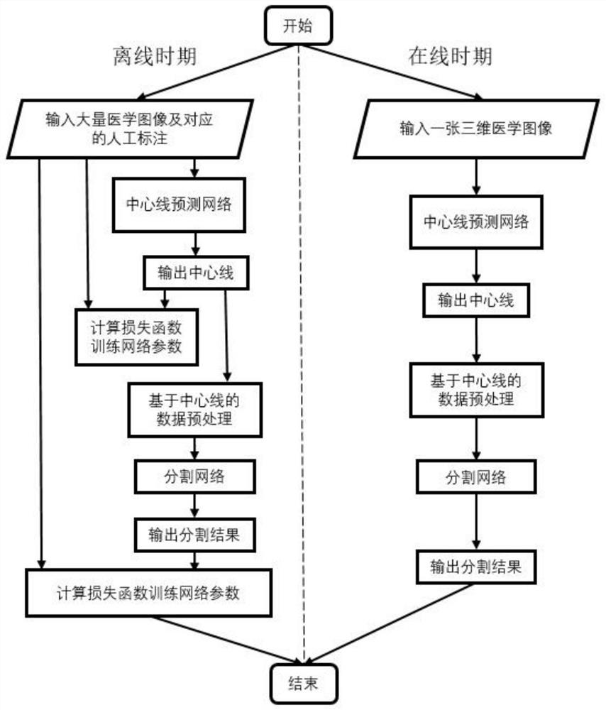
[0047] The present invention proposes a method for segmenting and marking multi-branched tubular structures in a three-dimensional image. The overall process is as follows: figure 1 As shown, the method is divided into an offline phase and an online phase, including the following steps:

[0048] 1) Offline stage;

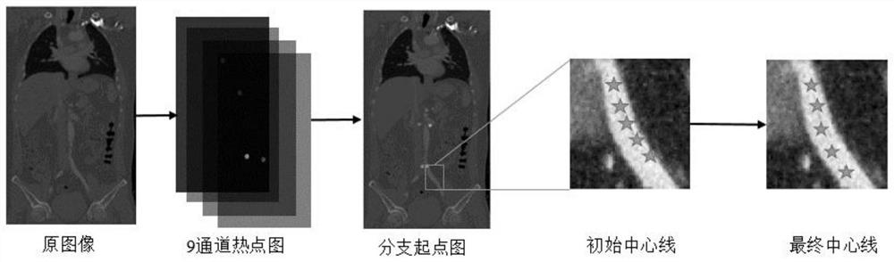
[0049] 1-1) Obtain the original image, mark the centerline and segmentation result of the tubular structure in each original image, and obtain the simple centerline training set and the complex centerline training set respectively; the specific steps are as follows:

[0050] 1-1-1) Obtain about 50 enhanced CT images of the same part as the original images, such as coronary artery problems need ...


PUM


Abstract

The invention provides a method for segmenting and marking a multi-branch tubular structure in a three-dimensional image, and belongs to the field of medical image processing. According to the method,a multi-branch tubular structure is regarded as a set of a plurality of single-branch tubular structures, and for the single-branch tubular structures, a center line is obtained through key point detection and neural network fine adjustment methods; then a cross section image of the tubular structure is acquired along the center line based on the center line, and finally a segmentation result ofthe tubular structure is acquired by using a segmentation network. The specific multi-branch tubular structure existing in the image can be segmented and automatically marked, and especially for someimages with lesions, such as aortic dissection, aortic aneurysm and coronary artery stenosis images, a good segmentation result is achieved.

Description

technical field [0001] The invention belongs to the field of medical image processing, in particular to a method for segmenting and marking multi-branched tubular structures in a three-dimensional image. Background technique [0002] Computed tomography angiography is a mainstream method of medical imaging. Doctors can obtain three-dimensional tomographic images of specific parts of the human body through computer-aided tomography. In these three-dimensional images, important functional tissues such as arteries, veins, and trachea are mostly multi-branched tubular structures. By observing and analyzing these tubular structures, doctors can have a more intuitive and intuitive understanding of the patient's physical condition without surgery. fully understand. Take the aorta as an example. The aorta is the main artery that transports blood to all parts of the body. There are many important branches in the chest cavity and abdominal cavity, which are responsible for delivering...

Claims


Application Information

Patent Timeline
Patent Type & Authority Applications(China)
IPC IPC(8): G06T7/00G06T7/12G06T7/60
CPCG06T7/0012G06T7/12G06T7/60G06T2207/10081G06T2207/30101G06T2207/10012G06T2207/20081
Inventor 冯建江周杰方辉谭子萌
Owner TSINGHUA UNIV
Why Eureka
Try Eureka