Method, device and equipment for segmenting ventricular region in brain CT image

A CT image and region segmentation technology, applied in the field of image processing, can solve problems such as inaccurate ventricle regions, achieve the effect of narrowing the image range and improving accuracy

Pending Publication Date: 2021-03-19
沈阳东软智能医疗科技研究院有限公司
View PDF0 Cites 2 Cited by
  • Summary
  • Abstract
  • Description
  • Claims
  • Application Information

AI Technical Summary

Problems solved by technology

Moreover, when segmenting the ventricle area in the brain CT image, it is easy to be affected by the imaging limitation of the brain CT image and the individual factors of the patient, resulting in inaccurate segmentation of the ventricle area.

Method used


Image

  • Method, device and equipment for segmenting ventricular region in brain CT image
  • Method, device and equipment for segmenting ventricular region in brain CT image
  • Method, device and equipment for segmenting ventricular region in brain CT image

Examples


specific Embodiment approach

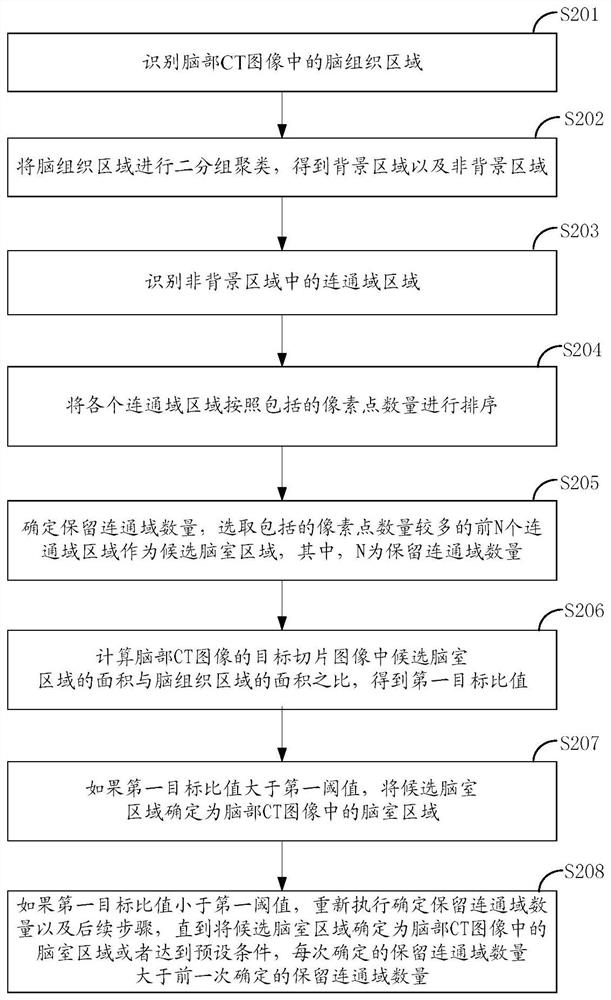
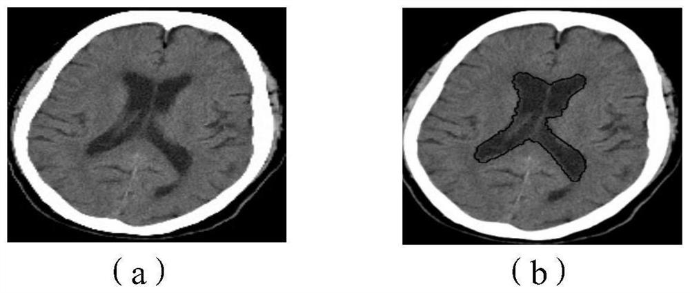
[0106] It is understandable that when composing candidate ventricular regions, regions with larger connected domains are preferentially selected. The embodiment of the present application provides a specific implementation method for determining the number of retained connected domains when each connected domain area is sorted according to the number of included pixels from large to small, specifically including:

[0107] Sequentially calculate the ratio of the number of pixels included in the two adjacent connected domain areas to obtain the second target ratio, stop the calculation when the second target ratio is greater than or equal to the second threshold, and sort the current two connected domain areas in The ranking value of the previous connected domain area is determined as the number of retained connected domains; the second threshold is increased with the number of times of performing the determination of the number of retained connected domains.

[0108] If the num...


PUM


Abstract

The embodiment of the invention discloses a method, device and equipment for segmenting a ventricular region in a brain CT image, and the method comprises the steps: recognizing a brain tissue regionin the brain CT image, carrying out the two-group clustering of the brain tissue region, and obtaining a background region and a non-background region of the brain tissue region; identifying connecteddomain areas in the non-background are, and sorting all the connected domain areas according to the number of included pixel points; determining the number of reserved connected domains, and selecting the first N connected domain regions including a large number of pixel points as candidate ventricular regions; judging whether the current candidate ventricular region can be determined as the ventricular region or not by utilizing the first target ratio and the first threshold value; and if not, adjusting the candidate ventricle. Therefore, the ventricular region obtained by segmentation is more complete and accurate. The ventricular region in the brain CT image can be automatically divided, so that the efficiency of dividing the ventricular region in the brain CT image is improved.

Description

technical field [0001] The present application relates to the field of image processing, and in particular to a method, device and equipment for segmenting ventricles in brain CT images. Background technique [0002] Brain CT scan is a common method for checking brain diseases. The brain CT images obtained by brain CT scan can be used to analyze the patient's condition. Among them, brain tissue image segmentation technology is an image processing technology for brain tissue segmentation of brain CT images. Through brain tissue segmentation technology, the size of each brain tissue and related information can be quickly determined, which is convenient for doctors to diagnose and diagnose diseases. treat. [0003] At present, the brain tissue image segmentation technology has strong pertinence, and manual auxiliary segmentation is required in the process of brain CT image segmentation. Moreover, when segmenting the ventricle area in the brain CT image, it is easy to be affec...

Claims


Application Information

Patent Timeline
Patent Type & Authority Applications(China)
IPC IPC(8): G06T7/11G06T7/194G06T7/136G06T7/62
CPCG06T2207/10081G06T2207/30016G06T7/11G06T7/136G06T7/194G06T7/62
Inventor 钱山袁红美杨明雷
Owner 沈阳东软智能医疗科技研究院有限公司
Why Eureka
Try Eureka