Supercharge Your Innovation With Domain-Expert AI Agents!

An image-assisted detection device for cardiovascular intubation

An auxiliary detection and cardiovascular technology, applied in hypnotic devices, devices that cause sleep or relaxation, and devices that cause changes in perception state, etc. and other problems to achieve the effect of facilitating detection, improving the effect, and avoiding waste.

Active Publication Date: 2021-11-23
新乡市中心医院
View PDF5 Cites 0 Cited by
  • Summary
  • Abstract
  • Description
  • Claims
  • Application Information

AI Technical Summary

Problems solved by technology

[0002] The heart is a hollow muscular organ located in the middle of the thoracic cavity. It is divided into two left and right chambers by a partition. Each chamber is divided into two parts: the upper atrium and the lower ventricle. The atrium collects blood from the heart, and the ventricle ejects The blood goes out of the heart, and the inlet and outlet of the ventricle have valves to ensure the one-way flow of blood. Especially when the heart is operated or detected, the doctor often needs to hold the laryngoscope assembly for a long time to detect; in the prior art, because the work The personnel need to hold the laryngoscope assembly and manipulate the laryngoscope assembly into the patient's body for a long time for observation, so the staff cannot avoid shaking their hands, making the laryngoscope assembly in the patient's body shake and collide with the pathogen in the patient's body, which affects observation on the one hand On the other hand, it will endanger the health of patients
[0003] In the prior art, there have also been some technical solutions on image-assisted detection devices for cardiovascular intubation. For example, a Chinese patent with application number CN202010839854.4 discloses an image-assisted detection device for cardiovascular intubation, including detection instruments and The laryngoscope component assembly, the monitoring instrument includes a display screen, an operation panel and a connecting line, and the tail end of the laryngoscope component component is electrically connected to the detection instrument through a connecting line; the technical solution is provided with an angle adjustment mechanism And the anti-slip mechanism, by rotating the limit sleeve 2, the distance between the limit sleeve 2 and the limit sleeve 1 is changed, which is convenient for the user to operate; but this technical solution does not completely solve the problem of the laryngoscope assembly caused by the shaking of the hands of the staff. The problem of shaking in the patient's body and contacting the pathogen will affect the staff's observation of the patient's condition and cause discomfort to the patient's body, which will cause the limitations of the technical solution

Method used


Image

  • An image-assisted detection device for cardiovascular intubation
  • An image-assisted detection device for cardiovascular intubation
  • An image-assisted detection device for cardiovascular intubation

Examples


Embodiment approach

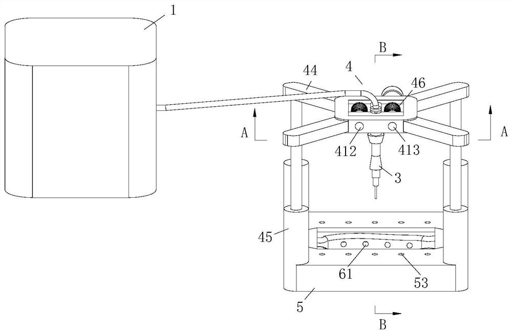
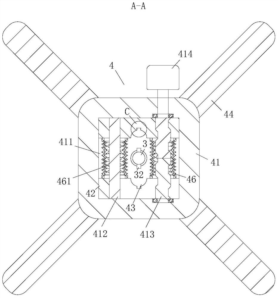
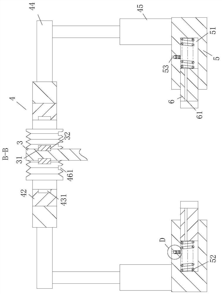
[0030] As an embodiment of the present invention, the surface of the tail end of the laryngoscope assembly 3 is provided with an annular groove 31; the bottom surface of the semicircular groove 43 is provided with a No. 1 groove 431; The groove 431 is at the same horizontal position, and the ring groove 31 is rotatably connected with the protrusion 32; the shape of the No. 1 groove 431 is the same as that of the protrusion 32; Airbag 46; the bottom of the corrugated airbag 46 is provided with an air hole 461; when the two clamping blocks 42 approach, the tail end of the laryngoscope assembly 3 is located between the two semicircular grooves 43, by rotating the tail end of the laryngoscope assembly 3 The protrusion 32 on the top, so that the protrusion 32 is facing the No. 1 groove 431, so that the protrusion 32 can be snapped into the No. 1 groove 431; and the two clamping blocks 42 are close to each other; the corrugated airbag 46 is squeezed, The gas in the corrugated airbag...


PUM


Abstract

The invention belongs to the technical field of cardiovascular intubation image-assisted detection, and in particular relates to a cardiovascular intubation image-assisted detection device, which includes a detector, a laryngoscope assembly, a controller, and a clamping device; the clamping device is used for To clamp the laryngoscope assembly, the clamping device includes a mounting plate and a clamping block; a through groove is opened inside the mounting plate; the cross-sectional shape of the through groove is rectangular, and a sliding bar is arranged inside the through groove; the two sides of the sliding bar The end is fixedly connected with the two side walls of the through groove; through the clamping device, the controller controls the clamping device to limit the position of the laryngoscope assembly. The component shakes, thereby reducing the contact between the laryngoscope component and the pathogen in the patient’s body, and improving the safety of the patient. On the other hand, the staff can leave the tail end of the laryngoscope component with both hands, so as to concentrate on observing the pathogen in the patient’s body, thereby improving detection. Effect.

Description

technical field [0001] The invention belongs to the technical field of image-assisted detection of cardiovascular intubation, and in particular relates to an image-assisted detection device for cardiovascular intubation. Background technique [0002] The heart is a hollow muscular organ located in the middle of the thoracic cavity. It is divided into two left and right chambers by a partition. Each chamber is divided into two parts: the upper atrium and the lower ventricle. The atrium collects blood from the heart, and the ventricle ejects The blood goes out of the heart, and the inlet and outlet of the ventricle have valves to ensure the one-way flow of blood. Especially when the heart is operated or detected, the doctor often needs to hold the laryngoscope assembly for a long time to detect; in the prior art, because the work The personnel need to hold the laryngoscope assembly and manipulate the laryngoscope assembly into the patient's body for a long time for observation...

Claims


Application Information

Patent Timeline
Patent Type & Authority Patents(China)
IPC IPC(8): A61B1/267A61B1/04A61B1/00A61G13/12A61B90/14A61M35/00A61M21/02
CPCA61B1/00064A61B1/00131A61B1/00147A61B1/04A61B1/267A61G13/121A61G13/126A61G2210/00A61M21/02A61M2021/0077A61M2210/0606A61B90/14A61M35/20A61M2210/005
Inventor 刘振
Owner 新乡市中心医院
Features
  • R&D
  • Intellectual Property
  • Life Sciences
  • Materials
  • Tech Scout
  • Unparalleled Data Quality
  • Higher Quality Content
  • 60% Fewer Hallucinations
Learn More