

Application of succinic acid in diagnosis or treatment of spontaneous abortion
A technology for spontaneous abortion and succinic acid, which is applied to medical preparations, pharmaceutical formulations, and drug combinations containing active ingredients, can solve problems such as no reports of succinic acid diagnosis or treatment of spontaneous abortion, and can reduce the level of succinic acid, embryonic Increased absorption rate, proliferative and invasive effects
- Summary
- Abstract
- Description
- Claims
- Application Information
AI Technical Summary
Problems solved by technology
Method used
Image

Examples
Embodiment 1
[0032] Example 1 Analysis of the level of succinic acid in the villi and decidua of normal pregnant women and spontaneous abortion patients
[0033] The villus and decidua tissues of 30 pairs of normal pregnant women and patients with recurrent spontaneous abortion (RSA) were collected, and metabolic profiling was performed using nuclear magnetic resonance (NMR), and more than 95% of the samples were detected including glucose, amino acids, fatty acids and choline metabolic pathways various types of metabolites. Among these metabolites, we found 27.5% lower succinate levels, 22.4% higher fumaric acid levels, and 21.1% lower lactate levels in the villi of RSA patients than controls (Table 1). However, the levels of each metabolite in decidual tissue were not statistically different (Table 2).
[0034] Table 1: Metabolite concentrations in the villi of normal pregnancy and spontaneous abortion patients
[0035]
[0036]
[0037] Note: Normal: villus tissue of normal preg...
Embodiment 2
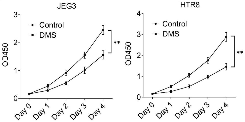
[0041] Example 2 Succinic acid promotes trophoblast proliferation and invasion
[0042] When treated with dimethyl succinate (DMS), JEG3 and HTR8 cells were plated at 2 × 10 3 The density of cells / well was placed in a 96-well plate (37°C, 5% CO 2 ), treated with 2 mM DMS for 24-96 hours. The medium was changed every day until the cells were harvested, then incubated with CCK-8 solution (C0039, Beyotime Biotechnology) for 2 h, and the absorbance was measured at 450 nm. The results show that DMS promotes trophoblast proliferation ( figure 1 ).
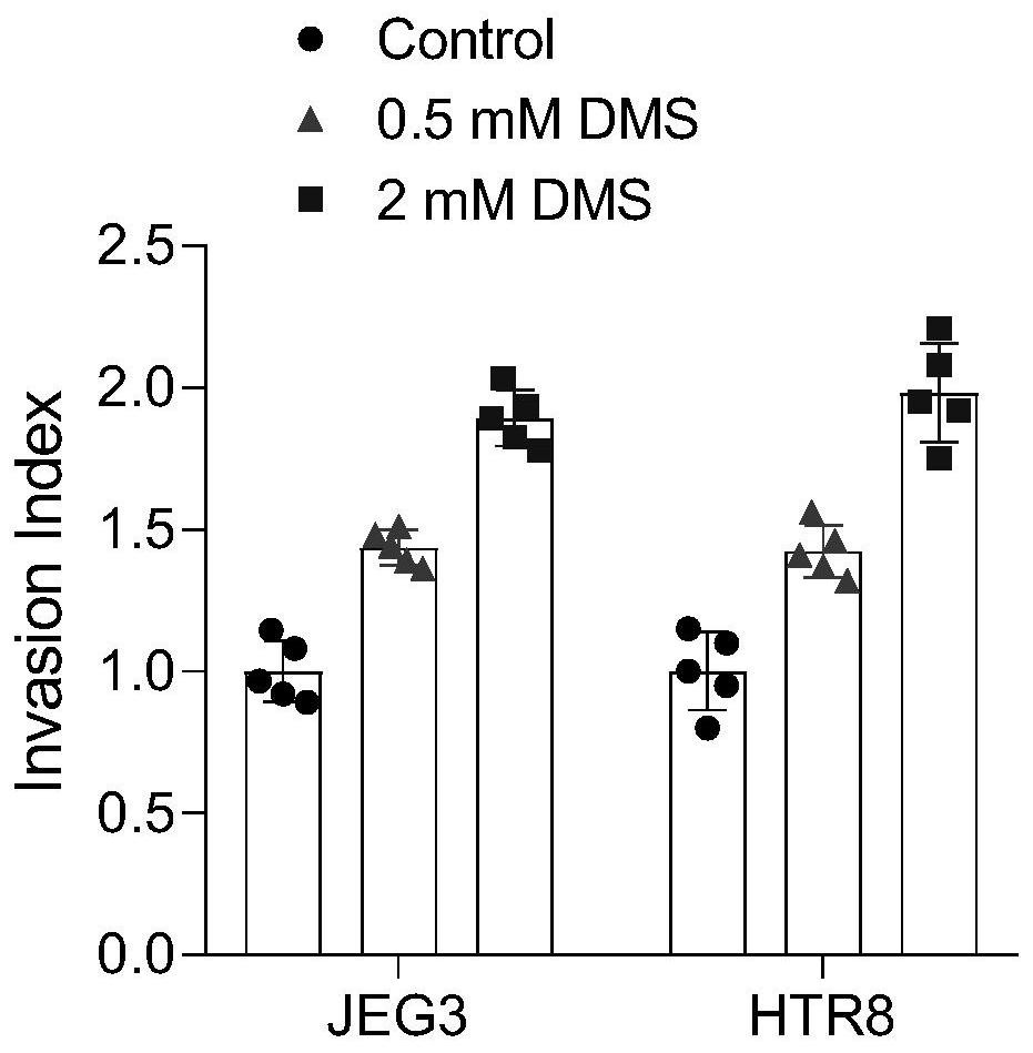
[0043] Invasion of JEG3 and HTR8 cells through Matrigel was objectively assessed by counting the number of cells in the invasion chamber that had migrated through the cell membrane. 15 μL of Matrigel (BD Biosciences) was pre-coated on a PET membrane (8 μm pore size, 6.5 mm diameter) on the upper surface of the filter of a Transewell plate (24-well plate, Corning, New York, USA) and air-dried under sterile conditions. Matrigel was tr...
Embodiment 3
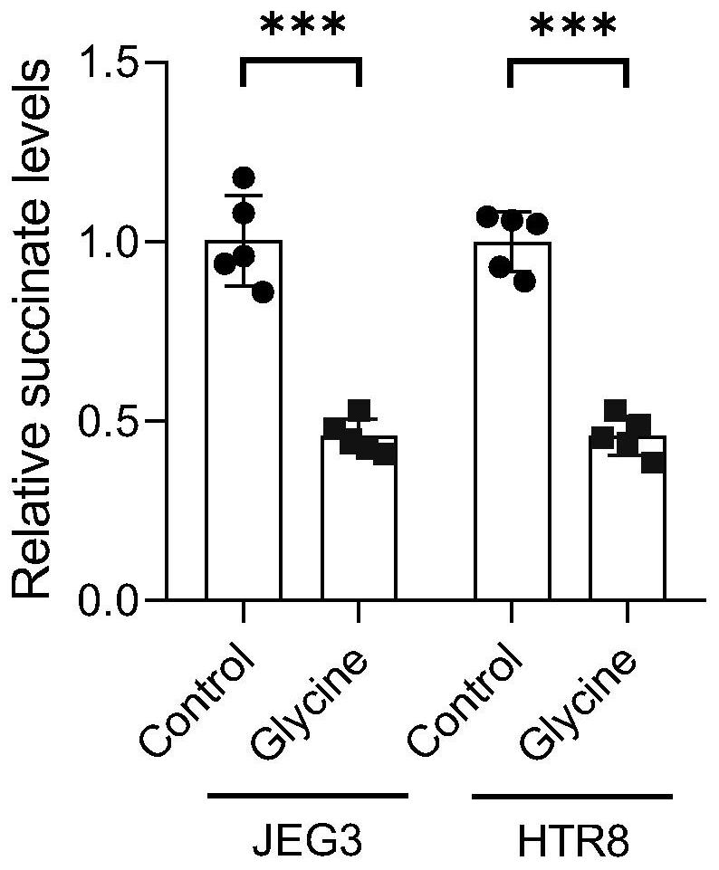
[0044] Example 3 Establishment of low succinate mouse model
[0045] Cellular experiments confirmed that glycine reduces succinate levels in trophoblast cell lines HTR8 and JEG3 ( image 3 ), intraperitoneal injection of glycine can reduce the level of succinate in mouse villus tissue ( Figure 4 ). In order to explore the effect of low succinic acid level on embryo implantation and spontaneous abortion, we selected 8-week-old female C57BL / 6 mice (weight 20-30 g, n=24) and randomly divided them into three groups: control group (intraperitoneal injection of normal saline), Glycine intraperitoneal injection group (glycine: 1500 mg / kg / day) and glycine+DMS intraperitoneal injection group (glycine: 1500 mg / kg / day+DMS: 3000 mg / kg / day). See vaginal suppository as 0.5 days of pregnancy, intraperitoneal injection from 0.5 days to 5.5 days, and the embryo absorption rate and the number of implanted embryos were counted on the 14.5th day. We found that intraperitoneal injection of gly...
PUM
 Abstract
Description
Claims
Application Information