Intravitreal tumor image result output system and method

A technology of tumor imaging and output methods, applied in neural learning methods, medical images, healthcare informatics, etc., can solve problems that affect clinicians' preoperative staging, complex and diverse tumor imaging performance, and affect curative effect

Active Publication Date: 2022-07-19
WEST CHINA HOSPITAL SICHUAN UNIV
View PDF5 Cites 0 Cited by
  • Summary
  • Abstract
  • Description
  • Claims
  • Application Information

AI Technical Summary

Problems solved by technology

The research results of CAD technology itself are not ideal, and many technical difficulties have yet to be overcome
[0003] 2) Eyeball tumors seriously endanger the life and visual function of the people. Among them, malignant tumors in the vitreous are the most common and serious types, and the delay in diagnosis and treatment brings huge economic pressure to the patient's family
Intravitreal tumor imaging has complex and diverse manifestations, and there is a lack of specialized imaging physicians. The interpretation of the images mainly relies on subjective experience, lacks standardized and standard report writing information, and there are also differences in report content and terminology. Accurately provided information on tumor staging is insufficient.
This affects the preoperative staging of clinicians, and the lack of interpretation of imaging studies by clinicians can easily lead to wrong staging before tumor treatment and affect the curative effect

Method used


Image

  • Intravitreal tumor image result output system and method
  • Intravitreal tumor image result output system and method
  • Intravitreal tumor image result output system and method

Examples


Embodiment 1

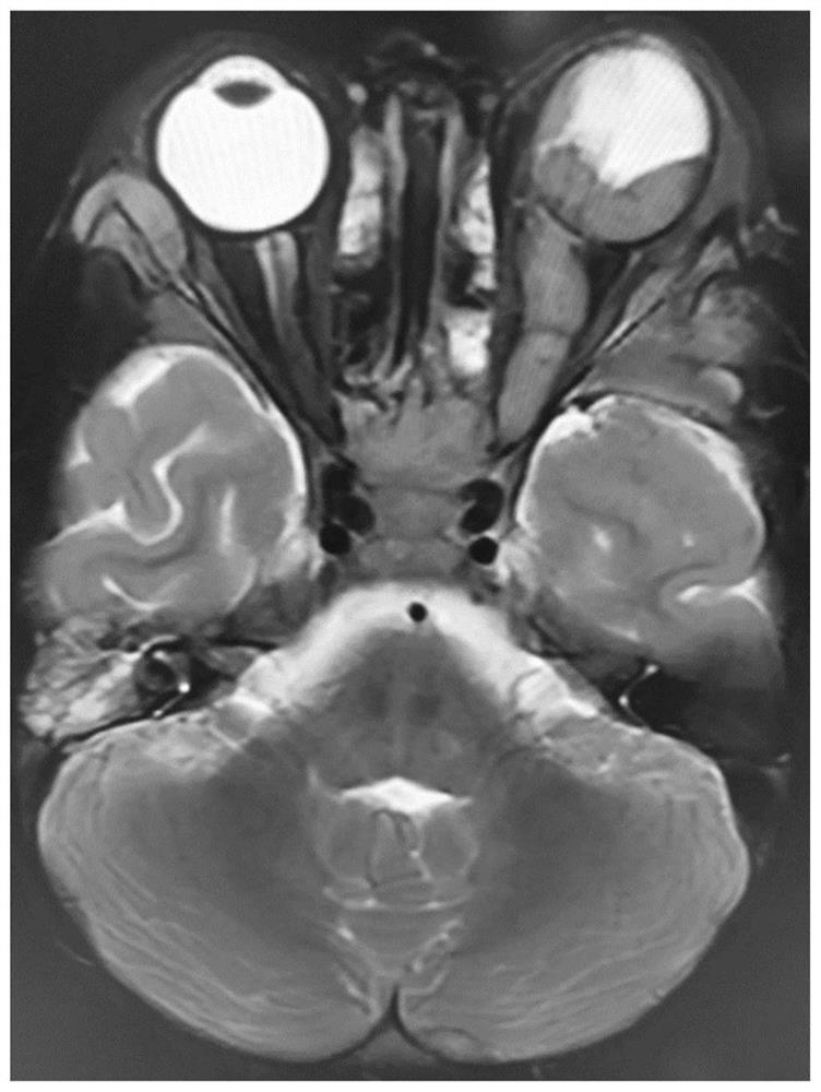
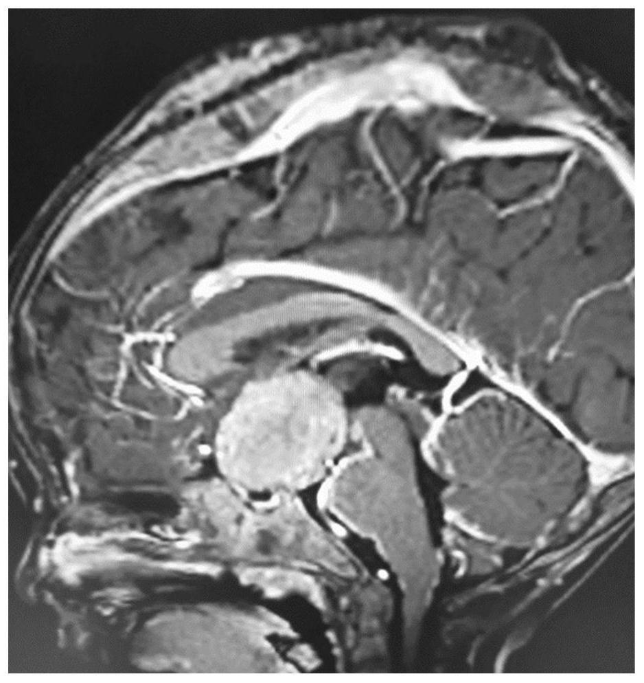
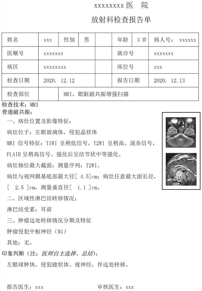
[0062] Basic structural model diagram of the orbit, including 6 extraocular muscles and intraocular anatomy. The extraocular muscles are skeletal muscles, and there are 4 rectus muscles and 2 oblique muscles that move the eyeball. The rectus muscle includes superior rectus, inferior rectus, medial rectus and lateral rectus, which together form the common tendon ring around the white optic canal, run forward along the wall of the eyeball, and insert at the upper, lower, medial, and upper parts of the sclera, respectively. Lateral; the obliques include the superior and inferior obliques. Intraocular structures include the anterior chamber, lens, ciliary body, macular optic disc, optic nerve, optic nerve head, and vitreous body.

[0063] In the first step, the imaging doctor judges the tumor site on the DICOM map, and clicks the site on the anatomical pattern map on the computer. The anatomical site can be the specific site of the tumor in the frame, such as the vitreous body, m...


PUM


Abstract

The present invention designs a visual lesion simulation map and a lesion map, which is convenient for dissection and comparison of the lesions. The signs observed by the radiologist are presented in a simple, standard, and easy-to-understand procedure. The method of clicking the patent map with the mouse avoids the error of manual input on the keyboard and the misinterpretation of the anatomical structure, realizes the standardization of image report terminology, the simplification of the operation interface, and the accurate and standardized description of the lesions. By comparing the lesion description result with the preset data module, the output of the description result is completed.

Description

technical field [0001] The present application relates to the field of MRI image acquisition and interpretation, and in particular, to a system and method for outputting image results of intravitreal tumors. Background technique [0002] 1) In 1959, American scholars Ledley et al. introduced mathematical models into clinical medicine for the first time, and applied them experimentally to a group of lung cancer cases. In 1966, Ledley innovatively proposed the concept of "computer assistance", which opened the prelude to the combination of computer and medicine. The research results of CAD technology itself are not ideal, and many technical difficulties remain to be overcome. [0003] 2) Tumors of the eyeball seriously endanger people's life and visual function. Among them, intravitreal malignant tumors are the most common and most serious type, and the delay in diagnosis and treatment brings huge economic pressure to the patient's family. At present, the main treatment meth...

Claims


Application Information

Patent Timeline
Patent Type & Authority Patents(China)
IPC IPC(8): G16H30/20G16H30/40G06N3/08G06N3/04
CPCG16H30/20G16H30/40G06N3/084G06N3/045
Inventor 杨喜彪王哲涛冷琦
Owner WEST CHINA HOSPITAL SICHUAN UNIV
Why Eureka
Try Eureka