
Probe integrating optical coherence tomography and intravascular ultrasound

A technology of coherence tomography and integrated optics, applied in the field of diagnosis, can solve problems such as increasing the incidence of device-related complications and increasing the economic burden of patients

Active Publication Date: 2021-08-10
HARBIN MEDICAL UNIVERSITY +1
View PDF8 Cites 1 Cited by
  • Summary
  • Abstract
  • Description
  • Claims
  • Application Information

AI Technical Summary

Problems solved by technology

In clinical diagnosis and treatment, the detailed information of lesions helps to better evaluate plaque stability and formulate personalized diagnosis and treatment strategies. Invasive imaging increases the incidence of device-related complications and increases the financial burden on patients

Method used


Image

  • Probe integrating optical coherence tomography and intravascular ultrasound
  • Probe integrating optical coherence tomography and intravascular ultrasound
  • Probe integrating optical coherence tomography and intravascular ultrasound

Examples


Embodiment Construction

[0025] The present invention will be further described in detail below in conjunction with the accompanying drawings.

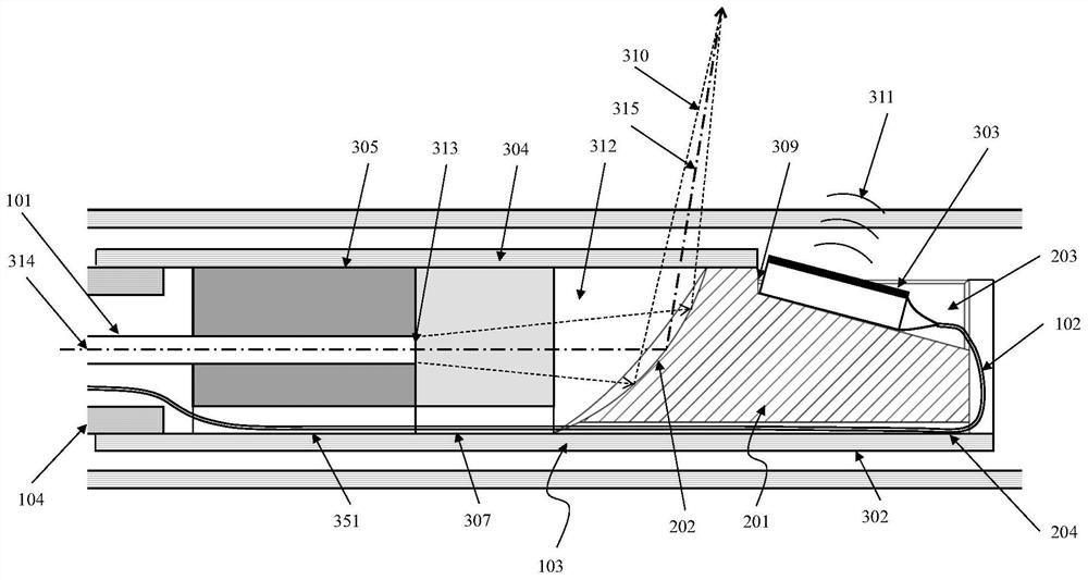
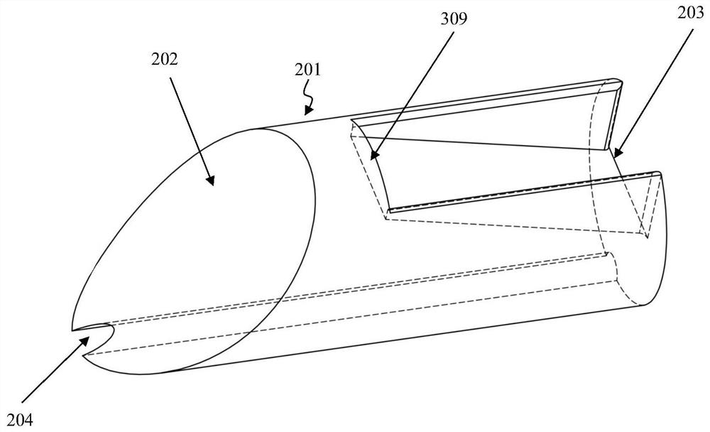
[0026] Such as figure 1 The illustrated embodiment of the present invention includes: a catheter 100, an integrated probe 103 and a spring tube 104, wherein the catheter 100 is composed of a proximal catheter 107 at the proximal end, a telescopic part 106 at the middle and a distal catheter 105 at the distal end, wherein the telescopic The two ends of the part 106 are integrally connected with the proximal catheter 107 and the distal catheter 105 respectively, and the far and near ends of the telescopic part 106 can ensure relative axial sliding and circumferential rotation on the basis of keeping the seal, and the sleeve in the integrated probe 103 The tube 302 is affixed to the distal end of the spring tube 104, and the proximal end of the spring tube 104 is fixed in the proximal guide tube 107 through a snap groove and a snap ring, thereby ensuring that th...


PUM

PropertyMeasurementUnit
The inside diameter ofaaaaaaaaaa
Wall thicknessaaaaaaaaaa
Axial lengthaaaaaaaaaa

Abstract

The invention discloses a probe integrating optical coherence tomography and intravascular ultrasound, and belongs to the field of diagnosis. The probe comprises a catheter, an integrated probe and a bourdon tube, wherein the near end of a sleeve in the integrated probe is fixedly connected with the bourdon tube; an optical fiber connector, a spacer block and a reflector sensor integrated frame are sequentially and fixedly connected into the sleeve from the near end to the far end; an optical fiber enters the optical fiber connector from the near end of the sleeve and is used for centering the optical fiber; the optical fiber end face of the optical fiber is attached to and fixedly connected with the surface of the near end of the spacer block, the near end of the optical fiber is connected with an optical fiber connector in the patient interaction unit, and an ultrasonic probe installed in the reflector sensor integrated frame is connected with an ultrasonic connector in the patient interaction unit through a wire. According to the invention, the relative positions of the OCT probe and the IVUS sensor are consistent during batch production, so the registration of the two images is ensured. And the problem that the relative positions are difficult to directly match due to different acousto-optic emission and collection modes is solved through the independent reflector and sensor integrated frame.

Description

technical field [0001] The invention belongs to the technical field of diagnosis, and specifically relates to a probe integrating optical coherence tomography (OCT) and intravascular ultrasound (IVUS). state imaging. Background technique [0002] Intravascular imaging has been widely used in atherosclerotic diseases, especially coronary heart disease. The most commonly used imaging systems are intravascular ultrasound (IVUS) and optical coherence tomography (OCT). Both of them use intracoronary imaging catheters to image the cross-section of coronary arteries, and their different imaging principles make them have different imaging advantages. Since the penetrating ability of IVUS to tissue is better than that of OCT, it can realize the identification of the thickness of blood vessel wall, while the axial resolution of OCT is 10 times that of IVUS, which can realize the identification of plaque microstructure. OCT uses infrared light to image the lumen, and red blood cells ...

Claims


Application Information

Patent Timeline
IPC IPC(8): A61B8/12A61B8/08A61B5/00
CPCA61B8/12A61B8/0891A61B8/4444A61B8/5261A61B5/0066A61B5/0084
Inventor 于波康维候静波贾海波赵晨徐晨阳
Owner HARBIN MEDICAL UNIVERSITY
Learn More