Intestinal lesion auxiliary diagnosis method based on non-normalized depth residual error and attention mechanism
A technology for assisting diagnosis and attention, applied in the field of medical image processing, can solve the problems of blurred boundary walls of lesions, inadequate extraction of subtle features, and large differences in the size and shape of intra-class lesions, so as to overcome large differences in shape and improve classification performance. Effect
- Summary
- Abstract
- Description
- Claims
- Application Information
AI Technical Summary
Problems solved by technology
Method used
Image

Examples
Embodiment Construction
[0030] The present invention will be further described below in conjunction with the accompanying drawings.
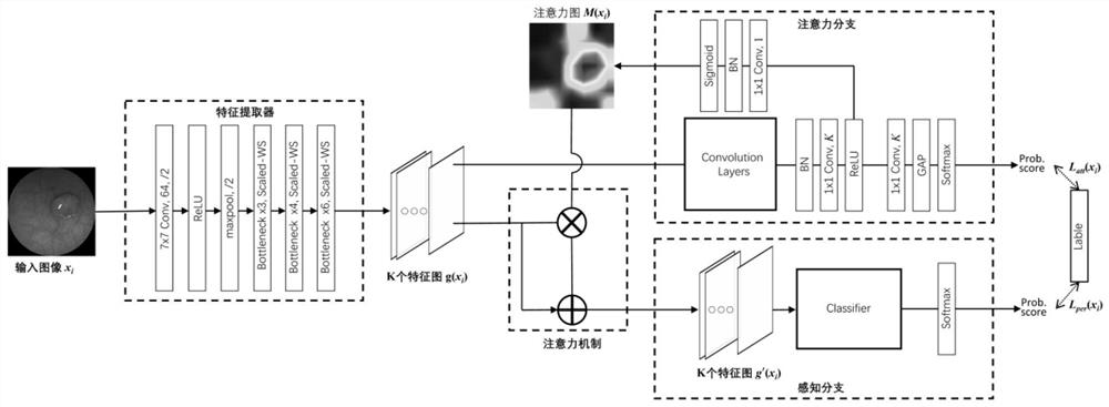
[0031] refer to Figure 1 to Figure 5 , a method for auxiliary diagnosis of intestinal lesions based on unnormalized depth residual and attention mechanism, comprising the following steps:
[0032] Step 1: Input image dataset X={x 1 ,x 2 ,...,x n}, where the X matrix represents the data set, n represents the total sample size, and x i ∈ R 224×224×3 Represents the feature vector composed of three channel pixel values of the input image, (x i ,y i ) means sample i, y i Indicates the sample category label, its value is 0 for normal, its value is 1 for polyps, its value is 2 for ulcers, when a classification model is trained, the image feature vector x i As input, predict whether the output result label is 0, 1 or 2, so that it can be judged whether the picture is normal, has polyps or has ulcers;
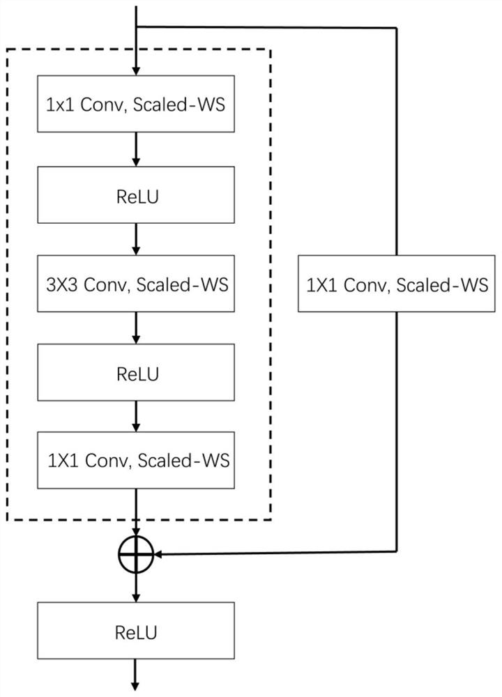
[0033] Step 2: Due to the large differences in the size and sha...
PUM
 Abstract
Description
Claims
Application Information
 - R&D
- Intellectual Property
- Life Sciences
- Materials
- Tech Scout
- Unparalleled Data Quality
- Higher Quality Content
- 60% Fewer Hallucinations
Browse by: Latest US Patents, China's latest patents, Technical Efficacy Thesaurus, Application Domain, Technology Topic, Popular Technical Reports.
© 2025 PatSnap. All rights reserved.Legal|Privacy policy|Modern Slavery Act Transparency Statement|Sitemap|About US| Contact US: help@patsnap.com