Stacked projection reconstruction method for segmented small-area tumor blocks

A small area, tumor technology, applied in the field of graphics and computer vision, can solve the problem of inapplicability, and achieve the effect of overcoming the volume effect

Active Publication Date: 2021-08-13
NANJING UNIV OF INFORMATION SCI & TECH
View PDF4 Cites 2 Cited by
  • Summary
  • Abstract
  • Description
  • Claims
  • Application Information

AI Technical Summary

Problems solved by technology

[0004] Aiming at the deficiencies of the existing medical diagnosis technology, the present invention provides a stacking projection reconstruction method of small-area tumor blocks after segmentation. After reconstruction, the texture information can accurately locate partitions and volumes intuitively, and reversely avoids the problem of inapplicability of specific parts.

Method used


Image

  • Stacked projection reconstruction method for segmented small-area tumor blocks
  • Stacked projection reconstruction method for segmented small-area tumor blocks
  • Stacked projection reconstruction method for segmented small-area tumor blocks

Examples


Embodiment Construction

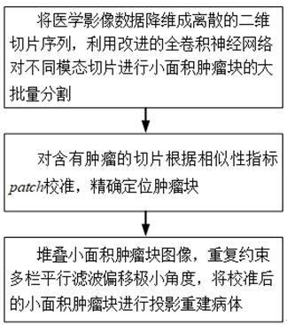
[0034] Aiming at medical imaging of different modalities of all organs such as brain, lung, heart, etc., the present invention provides a method for stacking projection reconstruction of a segmented small-area tumor block, which includes the following steps:

[0035] Step 1. Reduce the dimensionality of medical imaging data into discrete two-dimensional slice sequences, and use the improved fully convolutional neural network to perform large-scale segmentation of small-area tumor blocks on different modality slices;

[0036] Among them, reducing the dimensionality of medical image data into discrete two-dimensional slice groups includes the following steps:

[0037] Step 1-1, to avoid tumor site specificity, combine the built-in Slice() and Crop() to slice the pixel map of each case orthogonally from the center of gravity to form m sequence of slices, m The value is determined by the quality of clinical medical images to ensure the validity and reliability of reconstruction; ...


PUM


Abstract

The invention provides a stacked projection reconstruction method for small-area tumor blocks after segmentation. The method comprises the following steps: step 1, carrying out dimensionality reduction on medical image data to form a discrete two-dimensional slice sequence, and carrying out large-scale segmentation of small-area tumor blocks on different modal slices by utilizing an improved full convolutional neural network; step 2, calibrating the slices containing the tumor according to a similarity index patch, and accurately positioning a tumor block; and step 3, stacking small-area tumor block images, repeatedly constraining multi-column parallel filtering to shift a minimum angle, and projecting the calibrated small-area tumor blocks to reconstruct a sick body. The objective of the method is to realize rapid segmentation, accurate positioning and reverse reconstruction of a sick body so as to display multi-dimensional structure information, display the distribution condition and the relative position of tumor tissues independently or in a combined manner, and promote the development of emerging medical schemes such as image navigation minimally invasive surgery and the like.

Description

technical field [0001] The invention belongs to the technical fields of graphics and computer vision, and in particular relates to a stacking projection reconstruction method of a segmented small-area tumor block. Background technique [0002] Medical imaging technology refers to the analysis and processing of patient image data through advanced computer software and hardware, which has become an important part of medical information construction. Among them, imaging technologies such as computed tomography (CT), magnetic resonance imaging (magnetic resonance imaging, MR), and digital radiography (digital radiography, DR) have a great impact on the diagnosis and treatment of tumors. value. The medical visualization interface can allow doctors to instruct and introduce relevant image information to patients in detail, intuitively and accurately understand the size, location and morphological characteristics of the entire lesion area, thereby improving the objectivity of clin...

Claims


Application Information

Patent Timeline
Patent Type & Authority Applications(China)
IPC IPC(8): G06T11/00G06T7/10G06T7/70G06T5/00G06F3/0484G06N3/04
CPCG06T11/003G06T7/10G06T7/70G06T5/002G06F3/04845G06T2207/30096G06N3/045
Inventor 谈玲马雯杰夏景明
Owner NANJING UNIV OF INFORMATION SCI & TECH
Why Eureka
Try Eureka