

A Stacked Projection Reconstruction Method for Small-area Tumor Blocks after Segmentation
A small area, tumor technology, applied in the field of graphics and computer vision, can solve the problem of inapplicability, and achieve the effect of overcoming the volume effect
- Summary
- Abstract
- Description
- Claims
- Application Information
AI Technical Summary
Problems solved by technology
Method used
Image

Examples
Embodiment Construction
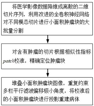
[0030] Aiming at medical imaging of different modalities of all organs such as brain, lung, heart, etc., the present invention provides a method for stacking projection reconstruction of a segmented small-area tumor block, which includes the following steps:
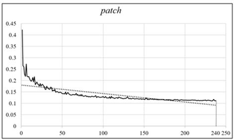
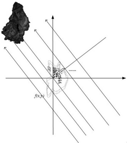
[0031] Step 1. Reduce the dimensionality of medical imaging data into discrete two-dimensional slice sequences, and use the improved fully convolutional neural network to perform large-scale segmentation of small-area tumor blocks on different modality slices;
[0032] Among them, reducing the dimensionality of medical image data into discrete two-dimensional slice groups includes the following steps:
[0033] Step 1-1, in order to avoid tumor site specificity, combine the built-in Slice() and Crop() to slice the pixel map of each case orthogonally from the center of gravity to form m sequence of slices, m The value is determined by the quality of clinical medical images to ensure the validity and reliability of reconst...
PUM
 Abstract
Description
Claims
Application Information