
Magnetic resonance-based method and system for determination of oxygen saturation in flowing blood

a magnetic resonance and blood flow technology, applied in the field of magnetic resonance techniques for differentiation of tissue signals, can solve the problems of low spatial resolution, affecting measurement accuracy, and particularly challenging, and achieve the effect of improving spatial resolution and overcomplicating partial volume effects

Inactive Publication Date: 2016-04-12
HOSPITAL FOR SICK CHILDREN +1
View PDF5 Cites 0 Cited by
  • Summary
  • Abstract
  • Description
  • Claims
  • Application Information

AI Technical Summary

Benefits of technology

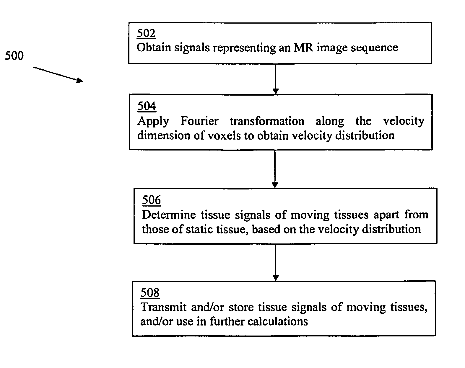
The patent describes a method and system for usingMR technology to determine the movement of blood within vessels and the surrounding tissue. This helps to separate the signals from the moving tissue and the surrounding static tissue, which can be useful for determining blood oxygen saturation. The technique involves analyzing the velocity dimension of an MR data set, which can help to overcome partial volume effects without needing to increase the resolution of the data.

Problems solved by technology

This may be particularly challenging where spatial resolution is low.
MRI has been used for oximetry, but partial volume effects may affect the accuracy of the measurement.
However, conventional non-invasive oximetry methods may not be suitable in some cases, for example in premature babies with patent ductus arteriosus (PDA).
PDA is a heart problem that occurs soon after birth in some babies.
The invasive nature of this pulmonary artery oxygen measurement may be stressful for the patient, for example an already fragile infant.
Non-invasive techniques may be erroneous or may provide only indirect and / or very rough estimates of blood oxygen levels in such patients (e.g., limited to peripheral vessels).
However, this conventional MR oximetry technique may be unsuitable for accurate determination of oxygen saturation in relatively small blood vessels (e.g., vessels in infants and children).
As such, this technique has seen relatively little application to use in neonatal infant care.
This may be overcome by increasing the spatial resolution (e.g., using smaller image pixels or voxels), but this typically leads to a decrease in signal-to-noise of the measurement and, as a result, may produce less accurate T2 values.
Increasing the spatial resolution may also require a longer time for image acquisition, which may be unsuitable for very young patients or any other patients who are unable to hold still for a lengthy period of time.

Method used


Image

  • Magnetic resonance-based method and system for determination of oxygen saturation in flowing blood
  • Magnetic resonance-based method and system for determination of oxygen saturation in flowing blood
  • Magnetic resonance-based method and system for determination of oxygen saturation in flowing blood

Examples


examples

[0056]Examples of the disclosed method and system are now described. These examples are provided for the purpose of illustration only and are not intended to be limiting.

[0057]In one example, an example of the disclosed method and system is used to calculate T2 and oxygen saturation in a phantom. The disclosed method is compared with a conventional method.

[0058]In the phantom, to mimic blood flowing in a vessel, water was pumped through a thin-walled (e.g., thickness=0.5 mm) latex tube at a constant flow rate by a computer-controlled gear pump. The tube was immersed in a water bath to mimic static tissue surrounding the vessel and a contrast agent, in this example gadopentetate dimeglumine (e.g., Magnevist™, Berlex, Canada) was used to adjust the tubing and bath T2 values to represent arterial blood and tissue, respectively. Imaging was performed on a 1.5 T MR system (GE Healthcare, USA) using the body coil.

[0059]To consider the suitability of the disclosed method in the presence of...


PUM


Abstract

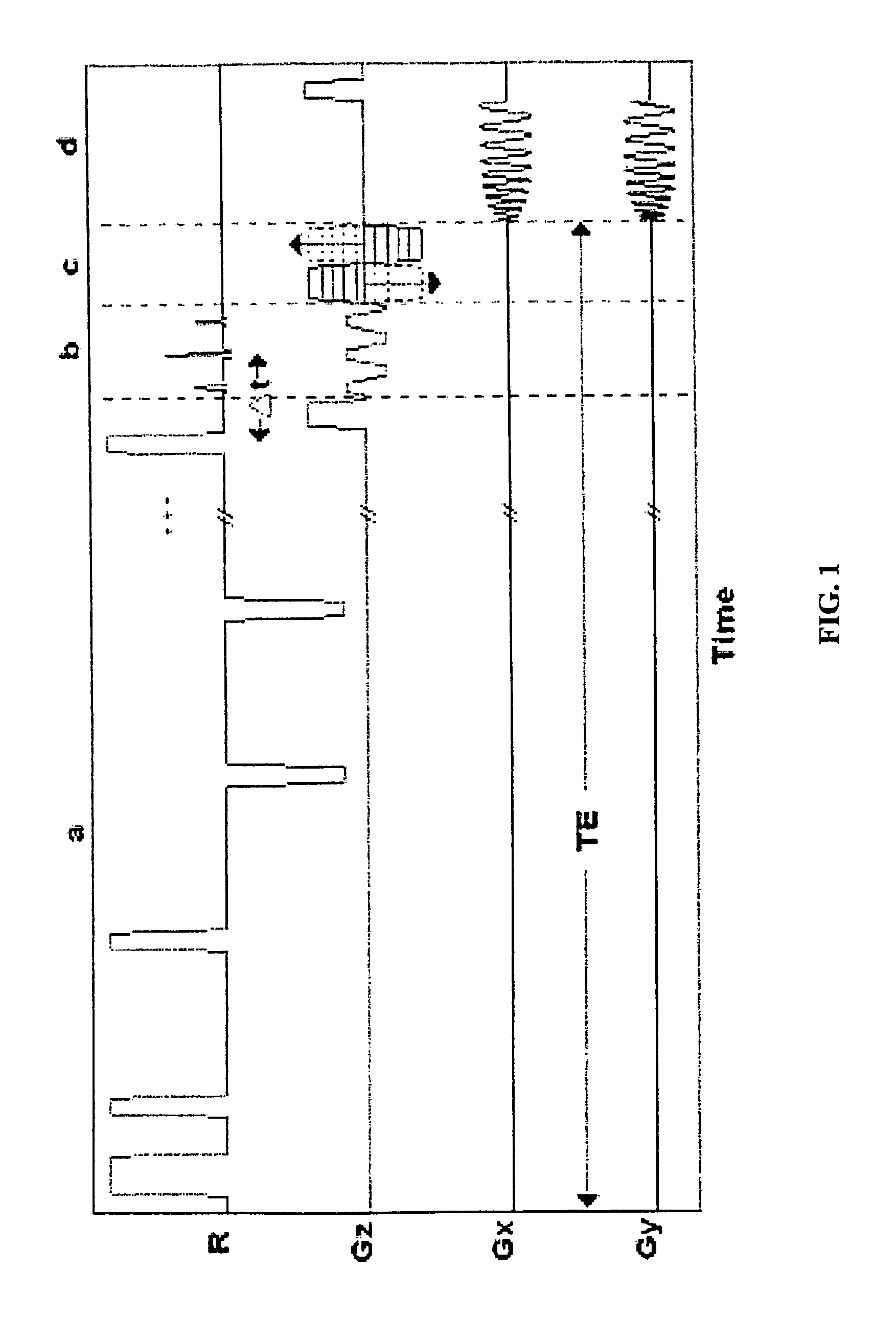
A method and system for determination of oxygen saturation in blood flowing in a vessel using magnetic resonance (MR). An MR image sequence is acquired with different echo time (TE) encoding, and different Fourier velocity encoding (FVE). A Fourier transformation is applied along the velocity dimension to determine a velocity distribution of tissue signals in each voxel of the image sequence. Tissue signals indicative of moving tissues are separated from tissue signals indicative of static tissue, based on the velocity distribution. Oxygen saturation in blood may then be determined using only the tissue signals indicative of flowing blood.

Description

CROSS-REFERENCE TO RELATED APPLICATION[0001]The present disclosure claims priority from U.S. provisional patent application No. 61 / 324,807, filed Apr. 16, 2010, the entirety of which is hereby incorporated by reference.TECHNICAL FIELD[0002]The present disclosure relates generally to magnetic resonance techniques for differentiation of tissue signals. In particular, the present disclosure may be useful for determination of oxygen saturation in blood flowing in a vessel.BACKGROUND[0003]In magnetic resonance imaging (MRI), signals obtained from moving tissues (e.g., flowing blood) may be contaminated with signals from static tissues (e.g., vessel wall), in an example of partial volume effects. This may be particularly challenging where spatial resolution is low. MRI has been used for oximetry, but partial volume effects may affect the accuracy of the measurement.[0004]The use of oximetry is a well understood and recognized technique for the measurement of blood oxygen saturation levels...

Claims


Application Information

Patent Timeline
Patent Type & Authority Patents(United States)
IPC IPC(8): G01R33/483A61B5/026A61B5/145G01R33/28G01R33/565A61B5/00G01R33/50A61B5/11A61B5/02G01R33/563
CPCG01R33/483A61B5/0263A61B5/14542A61B5/7257G01R33/28G01R33/50G01R33/56509A61B5/02007A61B5/1128G01R33/56316
Inventor WERNIK, CHRISTOPHERSWAMINATHAN, VENKATWRIGHT, GRAHAMMACGOWAN, CHRISTOPHER
Owner HOSPITAL FOR SICK CHILDREN
Learn More