Three-dimensional lung cancer model support, preparation method and application
A lung cancer and model technology, applied in biochemical equipment and methods, respiratory/lung cells, tumor/cancer cells, etc., can solve the problems of large demand for decellularization reagents, poor mechanical properties of natural materials, and lack of flexibility in operation. Achieve the effects of accurately predicting the curative effect in vivo, good proliferation space, and good permeability of the stent
- Summary
- Abstract
- Description
- Claims
- Application Information
AI Technical Summary
Problems solved by technology
Method used
Image

Examples
Embodiment 1
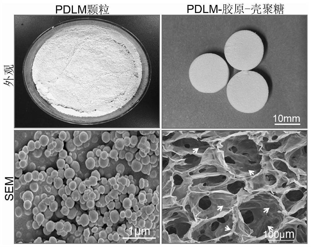
[0037] This embodiment provides a method for preparing a three-dimensional lung cancer model bracket, which specifically includes the following steps:
[0038] S1: Take pig-derived whole lung tissue, wash it with pure water for 3-5 times, completely remove the bronchial part, and grind the remaining part into 0.2cm 3 ~0.5cm 3 the crumbs;
[0039] S2: Wash the mince in step S1 with phosphate (PBS) buffer solution with a pH of 7.4 at room temperature for 3 to 5 times to remove the blood streaks. Stir with a magnetic stirrer for each wash. 0.5~1h;
[0040] S3: Add an aqueous solution of sodium dodecylsulfonate (SDS) with a mass fraction of 0.1-0.5% to the minced powder treated in step S2 at room temperature, stir with a magnetic stirrer for 5-10 hours, and initially remove cells, Partially decellularized lung fragments were obtained;
[0041] S4: Add a TritonX-100 solution with a volume fraction of 0.1-0.25% to the minced powder treated in step S3 at room temperature, stir wi...
Embodiment 2
[0053] In this example, the whole porcine lung tissue is excised from the bronchi, cut into small pieces, and crushed with a grinder, with a size of about 0.5cm 3 , put the minced pig lungs in a beaker, add 1000mL of ultrapure water, stir magnetically for 30min, then replace with new ultrapure water, repeat three times, add 1000mL of PBS to wash 3 times, magnetically stir for 30min each time, until there is no obvious After the blood color, add 1000mL of 0.5% (wt%, the same below) SDS solution, and after magnetic stirring for 5h, replace with a new 0.25% SDS solution and continue stirring for 5h to remove cells; then, add 0.25% (v%, the same below) Stir 1000mL of TritonX-100 solution for 5h to further remove remaining cells; after the cells are removed, add 1000mL of PBS to wash 5 times, each time lasting 30min, for washing the decellularization reagent. The decellularized powder was pre-frozen in a refrigerator at -20°C for 5 hours, and then dried in a freeze dryer at -80°C f...
Embodiment 3
[0062] In this example, the whole porcine lung tissue is excised from the bronchi, cut into small pieces, and crushed to a size of about 0.2 cm using a grinder. 3 ; After that, put the minced pig lungs in a beaker, then add 1000mL ultrapure water to the beaker, and replace with new ultrapure water after magnetic stirring for 30 minutes. , after no obvious blood color in the sample, add 0.25% (wt%, the same below) SDS solution 1000mL, after magnetic stirring for 3 hours, replace with new 0.1% SDS solution and continue stirring for 3 hours to remove cells. Subsequently, add 1000 mL of 0.1% (v%, the same below) TritonX-100 solution and stir for 8 hours to further remove the remaining cells. After the cells are removed, add 1000 mL of PBS to wash 3 times, each time lasting 1 hour, for washing the decellularization reagent . Then, the decellularized powder was pre-frozen in a refrigerator at -30°C for 10 hours, and then dried in a freeze dryer at -75°C for 15 hours to obtain a dri...
PUM
| Property | Measurement | Unit |
|---|---|---|
| Aperture | aaaaa | aaaaa |
| Aperture | aaaaa | aaaaa |
Abstract
Description
Claims
Application Information
 - R&D Engineer
- R&D Manager
- IP Professional
- Industry Leading Data Capabilities
- Powerful AI technology
- Patent DNA Extraction
Browse by: Latest US Patents, China's latest patents, Technical Efficacy Thesaurus, Application Domain, Technology Topic.
© 2024 PatSnap. All rights reserved.Legal|Privacy policy|Modern Slavery Act Transparency Statement|Sitemap