Liver tumor image segmentation model training method

An image segmentation and model training technology, applied in the field of medical image processing, can solve the problems of missing tumor characteristics, failing to improve the effectiveness of liver tumor segmentation, and difficult to capture small liver tumors

Pending Publication Date: 2021-09-03
北京精诊医疗科技有限公司
View PDF6 Cites 1 Cited by
  • Summary
  • Abstract
  • Description
  • Claims
  • Application Information

AI Technical Summary

Problems solved by technology

Although this method can segment the liver very well, it may miss the characteristic features of the tumor, which cannot improve the effectiveness of liver tumor segmentation, and it is difficult to better capture small tumors in the liver

Method used


Image

  • Liver tumor image segmentation model training method
  • Liver tumor image segmentation model training method

Examples


Embodiment Construction

[0017] Below in conjunction with accompanying drawing and embodiment, technical solution of the present invention is described further:

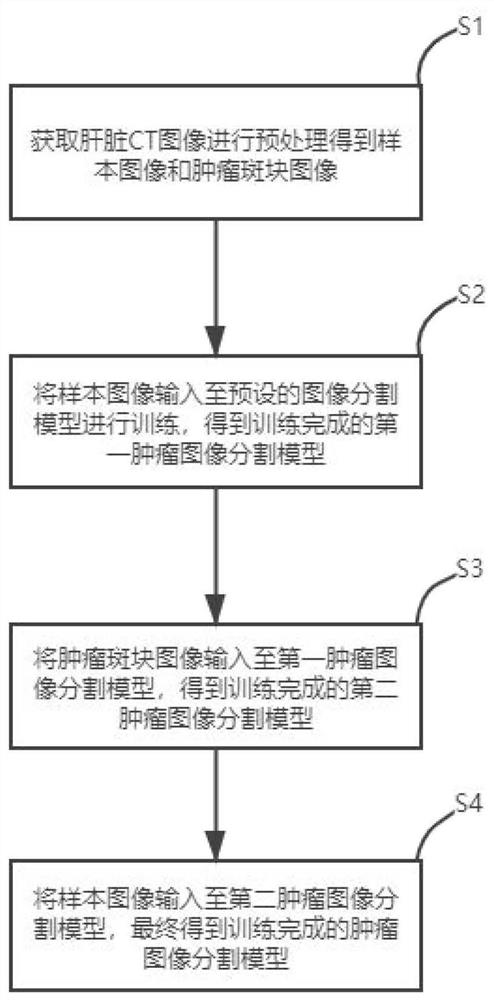
[0018] This embodiment provides a liver tumor image segmentation model training method, such as figure 1 shown, including:

[0019] Liver CT images are acquired, and the liver CT images include multiple groups of samples; in this embodiment, the training data includes 150 CT scans, all of which have a pixel resolution of 512×512.

[0020] Preprocessing operations are performed on liver CT images to obtain sample images and tumor plaque images. Preprocessing operations include interval interpolation, window transformation, effective range extraction and generation of tumor plaque images. In this embodiment, there are 12734 sample images in total, and the size is set to 64×256×256 to optimize the available GPU memory and the context information retained in the input patch.

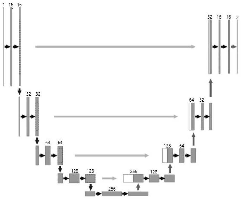
[0021] The tumor plaque image is used to encapsulate any tumor imag...


PUM


Abstract

The invention discloses a liver tumor image segmentation model training method, which comprises the following steps of: firstly, acquiring a liver CT (Computed Tomography) image, performing preprocessing operation on the liver CT image to obtain a sample image and a tumor plaque image, and then performing specific training on a tumor image segmentation model: at the first stage, inputting the sample image into a preset image segmentation model for training, obtaining a trained first tumor image segmentation model; in the second stage, inputting the tumor plaque image into the first tumor image segmentation model for training to acquire a second tumor image segmentation model after training is completed; and in the third stage, inputting the sample image into the second image segmentation model for training to acquire a tumor image segmentation model after training is completed. A network is trained from the two aspects of whole liver CT image input and tumor plaque image, the specific features of the tumor can be integrated, and the accuracy of liver tumor image segmentation is improved in combination with the overall background of the tumor data.

Description

technical field [0001] The invention belongs to the field of medical image processing, in particular to a method for training a liver tumor image segmentation model. Background technique [0002] Liver cancer is the second most common leading cause of cancer death worldwide. For liver cancer screening, computed tomography (CT) is the most commonly used imaging tool. Abnormal morphology and texture of the liver in CT, as well as visible lesions in primary and secondary It is an important marker of disease progression in recurrent liver neoplastic diseases. In clinical practice, the tumor size is much smaller than that of the whole CT as well as the size of the liver region, and segmenting tumors from the whole CT volume or liver region is a difficult challenge. [0003] In the prior art, a cascade model is usually used for liver tumor segmentation: liver localization network, liver segmentation network and tumor segmentation network. Although this method can segment the liv...

Claims


Application Information

Patent Timeline
Patent Type & Authority Applications(China)
IPC IPC(8): G06T7/11G06T7/00G06N3/08G06N3/04
CPCG06T7/11G06T7/0012G06N3/08G06T2207/20081G06T2207/20084G06T2207/30056G06T2207/30096G06T2207/10081G06N3/045
Inventor 王博赵威申建虎张伟徐正清
Owner 北京精诊医疗科技有限公司
Why Eureka
Try Eureka