
Weak supervision cervical cell image analysis method fusing global and local information

A technology of cervical cells and local information, applied in the field of cervical cytology pathological image analysis, can solve problems such as difficulty in obtaining

Active Publication Date: 2021-09-10
HEFEI UNIV OF TECH
View PDF6 Cites 4 Cited by
  • Summary
  • Abstract
  • Description
  • Claims
  • Application Information

AI Technical Summary

Problems solved by technology

[0005] In order to overcome the deficiencies of the prior art, the present invention proposes a weakly supervised cervical cell image analysis method that integrates global and local information, in order to quickly and accurately realize image classification and cell classification, and overcome the problem of detailed information labeling of pathological image data difficult to obtain

Method used


Image

  • Weak supervision cervical cell image analysis method fusing global and local information
  • Weak supervision cervical cell image analysis method fusing global and local information
  • Weak supervision cervical cell image analysis method fusing global and local information

Examples


Embodiment Construction

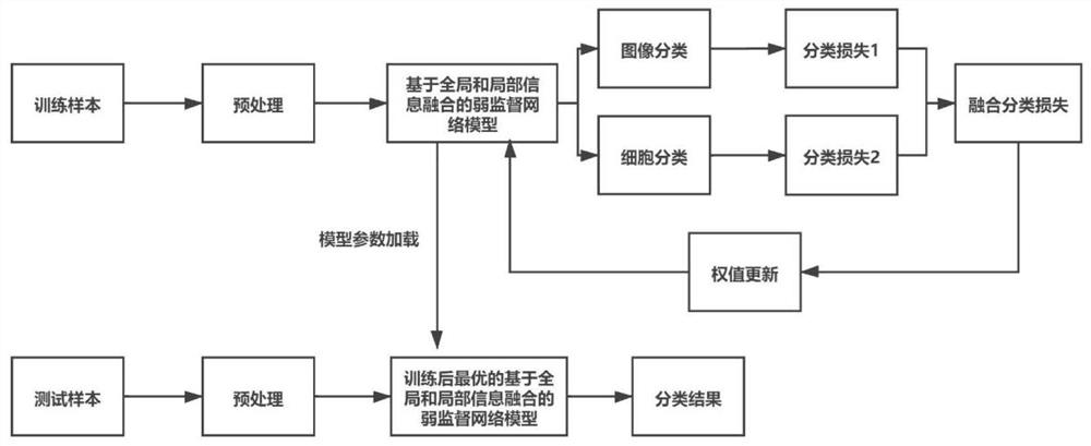
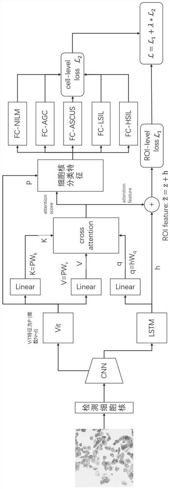
[0036] In this embodiment, a weakly supervised cervical cell image analysis method that integrates global and local information mainly uses a Vision Transformer (ViT) network structure and a bidirectional long-short-term memory network (BiLSTM) to extract the local and global images respectively. Information, and then use the cross-attention (CrossAttention) mechanism to fuse information of different scales to realize cervical liquid-based cell image classification and cell classification. Specifically, as figure 1 As shown, it is carried out according to the following steps:

[0037] Step 1. Obtain the cervical cell visual field image data set B={B 1 ,B 2 ,...,B n ,...,B N}, where B n Indicates the nth cervical cell view image containing several different types of cells, and the position and type of some cells are marked on the cervical cell view image; and there are: Indicates the cth cell in the nth cervical cell field of view image; C n Represents the total number o...


PUM


Abstract

The invention discloses a weak supervision cervical liquid-based cell image analysis method fusing global and local information. The method comprises the following steps: 1, acquiring a weak label cervical liquid-based cell view image data set with a picture level; 2, establishing a weak supervision network model fusing global and local information; 3, training a network model offline by using the data set of the weak label; and 4, predicting a cervical cell image by using the trained model, and completing image classification and detection and classification of a single cell. The defect that detailed labeling of cervical image data is limited can be overcome, and Field Of View (FOV)-level classification and cell-level detection and classification can be effectively completed by using a weak supervision method.

Description

technical field [0001] The invention relates to the field of cervical cytology pathological image analysis, in particular to a weakly supervised cervical liquid-based cell image analysis method that integrates global and local information. Background technique [0002] The traditional pathological image analysis method is as follows: a professionally trained pathologist needs to look at the pathological image under a microscope and find some regions of interest (Region Of Interest, ROI), and then the doctor will use his professional knowledge to classify and analyze. There are many problems in the traditional method. Doctors may need to read a large number of slides every day, and a single pathological slide image may contain millions of cells, which greatly increases the burden on doctors; The doctor's experience has a lot to do with it, which brings many difficulties to pathological data analysis. If the error is caused by the subjective opinion of the doctor who reads t...

Claims


Application Information

Patent Timeline
IPC IPC(8): G06K9/00G06K9/46G06K9/62G06N3/04G06N3/08
CPCG06N3/08G06N3/045G06F18/214
Inventor 史骏吴坤唐昆铭贺雨欣祝新宇孙宇李俊郑钰山姜志国
Owner HEFEI UNIV OF TECH
Learn More