
MRI brain tumor image segmentation method and system based on improved U-Net network

A brain tumor and image technology, applied in the field of image processing, can solve problems such as low-level feature redundant information, and achieve the effects of improving utilization, performance, and accuracy

Pending Publication Date: 2021-11-09
SHANDONG NORMAL UNIV
View PDF0 Cites 2 Cited by
  • Summary
  • Abstract
  • Description
  • Claims
  • Application Information

AI Technical Summary

Problems solved by technology

But the skip connection directly connects the atlas extracted by the encoder to the corresponding layer of the decoder, which leads to a large amount of redundant information in the low-level features;

Method used


Image

  • MRI brain tumor image segmentation method and system based on improved U-Net network
  • MRI brain tumor image segmentation method and system based on improved U-Net network
  • MRI brain tumor image segmentation method and system based on improved U-Net network

Examples


Embodiment 1

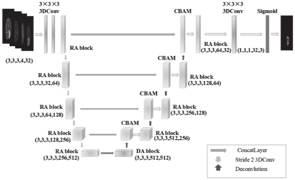
[0050] Such as figure 1 As shown, this embodiment provides a method for segmenting MRI brain tumor images based on the improved U-Net network. This embodiment uses this method as an example to illustrate the server. It can be understood that this method can also be applied to A terminal may also be applied to include a terminal, a server and a system, and may be realized through interaction between the terminal and the server. The server can be an independent physical server, or a server cluster or distributed system composed of multiple physical servers, or it can provide cloud services, cloud database, cloud computing, cloud function, cloud storage, network server, cloud communication, intermediate Cloud servers for basic cloud computing services such as software services, domain name services, security service CDN, and big data and artificial intelligence platforms. The terminal may be a smart phone, a tablet computer, a laptop computer, a desktop computer, a smart speaker...

Embodiment 2

[0069] This embodiment provides a system for segmenting MRI brain tumor images based on an improved U-Net network.

[0070] A segmentation system for MRI brain tumor images based on the improved U-Net network, including:

[0071] A segmentation module configured to: obtain the MRI brain tumor image to be segmented, input it into the trained improved U-Net network, and obtain an image marked with the segmented tumor;

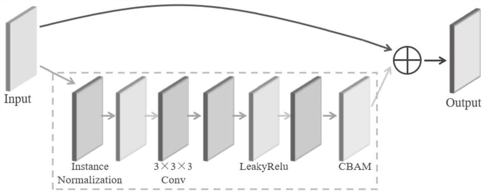
[0072] Model construction module, it is configured as: described improved U-Net network comprises: introduce the convolutional layer that replaces U-Net network with the residual module of double attention mechanism, introduce in U-Net network with attention The expansion pyramid module of the force mechanism, and introduces a double attention mechanism after each layer skip connection.

Embodiment 3

[0074] This embodiment provides a computer-readable storage medium on which a computer program is stored, and when the program is executed by a processor, the segmentation of MRI brain tumor images based on the improved U-Net network as described in the first embodiment is realized. steps in the method.


PUM


Abstract

The invention belongs to the technical field of image processing, and provides an MRI brain tumor image segmentation method and system based on an improved U-Net network. The method comprises the following steps: acquiring a to-be-segmented MRI brain tumor image, and inputting the to-be-segmented MRI brain tumor image into a trained improved U-Net network to obtain an image marked with a segmented tumor; and, according to the improved U-Net network, a residual module with a double-attention mechanism is introduced to replace a convolutional layer of the U-Net network, an expansion pyramid module with an attention mechanism is introduced into the U-Net network, and the double-attention mechanism is introduced after each layer skips connection.

Description

technical field [0001] The invention belongs to the technical field of image processing, in particular to a method and system for segmenting MRI brain tumor images based on an improved U-Net network. Background technique [0002] The statements in this section merely provide background information related to the present invention and do not necessarily constitute prior art. [0003] Brain tumors are unwanted, uncontrolled growths of cells in the human brain that can be classified as primary or secondary based on their origin. Primary brain tumors start in brain cells and spread to other parts of the brain. Secondary or metastatic tumors originate elsewhere in the body and spread to the brain. At present, the research on brain tumor segmentation mainly focuses on glioma. According to the World Health Organization (WHO) standard, glioma is divided into 4 grades, including grade I astrocytoma and grade II oligodendroglioma. , grade III inelastic glioma and grade IV glioma mu...

Claims


Application Information

Patent Timeline
IPC IPC(8): G06T7/11G06N3/04G06N3/08
CPCG06T7/11G06N3/084G06T2207/10088G06T2207/20016G06T2207/20081G06T2207/20084G06T2207/30096G06T2207/30016G06N3/045Y02T10/40
Inventor 王晶晶于子舒赵文瀚孙增钊李鸿祯张波赵蒙蒙刘建伟
Owner SHANDONG NORMAL UNIV
Learn More