Liver focus diagnosis method and device based on CT image

A technology of CT images and diagnostic methods, applied in the field of medical imaging, can solve the problems of difficulty in accurately diagnosing the type of lesions, inability to make full use of lesion information, etc., and achieve the effect of improving accuracy

Pending Publication Date: 2021-11-16
HANGZHOU SHENRUI BOLIAN TECH CO LTD +2
View PDF0 Cites 2 Cited by
  • Summary
  • Abstract
  • Description
  • Claims
  • Application Information

AI Technical Summary

Problems solved by technology

Diagnosis based on a single phase (such as plain scan) cannot make full use of lesion information in other phases, so it is difficult to accurately diagnose the type of lesion, especially in the case of the same shadow but different diseases

Method used


Image

  • Liver focus diagnosis method and device based on CT image
  • Liver focus diagnosis method and device based on CT image
  • Liver focus diagnosis method and device based on CT image

Examples


Embodiment Construction

[0055] In order to make the purpose, technical solution and advantages of the present invention clearer and clearer, the present invention will be further described below in conjunction with the accompanying drawings and specific embodiments. Apparently, the described embodiments are only some of the embodiments of the present invention, but not all of them. Based on the embodiments of the present invention, all other embodiments obtained by persons of ordinary skill in the art without making creative efforts belong to the protection scope of the present invention.

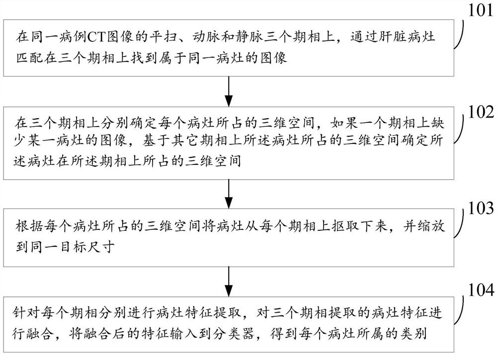
[0056] figure 1 It is a flowchart of a method for diagnosing liver lesions based on CT images according to an embodiment of the present invention, including the following steps:

[0057] Step 101, in the three phases of plain scan, artery and vein of the CT image of the same case, find images belonging to the same lesion in the three phases through liver lesion matching;

[0058]Step 102: Determine the three-dim...


PUM


Abstract

The invention provides a liver focus diagnosis method and device based on a CT image. The method comprises the following steps: finding an image belonging to the same lesion in three phases through liver lesion matching on the three phases of plain scanning, artery and vein of a CT image of the same case; respectively determining a three-dimensional space occupied by each focus on three phases, and if one phase lacks an image of a certain focus, determining a three-dimensional space occupied by the focus on the phase based on the three-dimensional spaces occupied by the focus on other phases; digging the focus down from each phase and scaling to the same target size; and respectively carrying out focus feature extraction on each phase, fusing the focus features extracted from the three phases, and inputting the fused features into a classifier to obtain the category to which each focus belongs. The invention can make full use of the expressions and signs of the lesion in multiple phases for lesion diagnosis, and improves the accuracy of lesion diagnosis.

Description

technical field [0001] The invention belongs to the technical field of medical imaging, in particular to a method and device for diagnosing liver lesions based on CT images. Background technique [0002] In recent years, with the development of deep learning and medical imaging technology, applying deep learning technology to medical images has become a relatively hot research field. At present, deep learning technology has achieved good results in related image processing of lungs, breasts and brains. [0003] A complete CT image contains at least three phases of plain scan, arterial and venous data. At present, the diagnostic technology of liver lesions using CT images is mainly based on plain CT scans to make a diagnosis of the nature of the lesions. Generally, the liver segmentation model is first used to obtain the segmented area of ​​the liver, and then the liver area on the plain scan is extracted as the input of the classification model, and the entire liver is dia...

Claims


Application Information

Patent Timeline
Patent Type & Authority Applications(China)
IPC IPC(8): G06T7/00G06T7/11G06K9/62G06T7/62G06T7/73G16H30/20G16H50/20
CPCG06T7/0012G06T7/11G06T7/62G06T7/73G16H50/20G16H30/20G06T2207/10081G06T2207/30056G06T2207/20081G06T2207/20084G06T2207/30096G06F18/22G06F18/241G06F18/253
Inventor 刘波王淑欣刘小青俞益洲李一鸣乔昕潘晶应汉宁蔡秀军
Owner HANGZHOU SHENRUI BOLIAN TECH CO LTD
Why Eureka
Try Eureka