
Medical image lesion position viewing method and system, equipment and storage medium

A medical imaging and storage medium technology, applied in the field of medical imaging image processing, can solve the problems of difficult and accurate comparison, error, time-consuming and labor-intensive, etc., and achieve the effect of improving efficiency and simplifying the operation of visual search and comparison.

Pending Publication Date: 2021-12-07
苏州复颖医疗科技有限公司
View PDF0 Cites 2 Cited by
  • Summary
  • Abstract
  • Description
  • Claims
  • Application Information

AI Technical Summary

Problems solved by technology

[0003] Many diseases require multiple imaging examinations to observe their progress and compare the results of multiple imaging. The existing model generally uses imaging plus imaging reports. In the imaging reports, the location and size of lesions are qualitatively described, and the previous inspections are reviewed. When comparing, it is necessary to visually search for comparisons based on the image report description in previous images, which is time-consuming and laborious
If the user has multiple lesions in one area, he or she will generally select a representative one or several for description in the image report, but it is difficult to make a one-to-one accurate comparison in the later stage, resulting in errors

Method used


Image

  • Medical image lesion position viewing method and system, equipment and storage medium
  • Medical image lesion position viewing method and system, equipment and storage medium
  • Medical image lesion position viewing method and system, equipment and storage medium

Examples


Embodiment approach

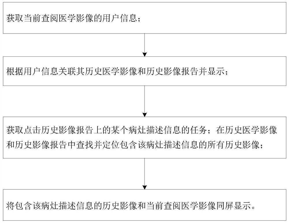
[0041] Such as figure 1 As shown, the first object of the present invention is to provide a method for checking the location of medical imaging lesions, including the following steps:

[0042] Obtain the information of users who are currently reviewing medical images;

[0043] Correlate and display historical medical images and historical image reports based on user information;

[0044] Obtain the task of clicking on a lesion description information on the historical image report; find and locate all historical images containing the lesion description information in the historical medical images and historical image reports;

[0045] The historical image containing the description information of the lesion and the currently consulted medical image are displayed on the same screen.

[0046] The marking and direct jumping method of the present invention simplifies the subsequent operation of visual search and comparison, enables accurate positioning of multiple lesions, and p...


PUM


Abstract

The invention provides a medical image lesion position viewing method and system, equipment and a storage medium. The method comprises the following steps: acquiring user information currently looking up a medical image; associating and displaying the historical medical image and the historical image report according to the user information; acquiring a task of clicking certain focus description information on the historical image report; searching and positioning all historical images containing the lesion description information in historical medical images and historical image reports; and displaying the historical image containing the focus description information and the current consulted medical image on the same screen.

Description

technical field [0001] The present invention relates to the field of medical imaging image processing, in particular to a method, system, device and storage medium for checking the location of medical imaging lesions. Background technique [0002] The existing digital medical images have all replaced analog images, and informatization-network has become an indispensable means and platform. Medical imaging informatization provides fast and accurate imaging diagnostic information for clinical practice. [0003] Many diseases require multiple imaging examinations to observe their progress and compare the results of multiple imaging. The existing model generally uses imaging plus imaging reports. In the imaging reports, the location and size of lesions are qualitatively described, and the previous inspections are reviewed. When comparing, it is necessary to visually search for comparisons based on the image report description in previous images, which is time-consuming and labo...

Claims


Application Information

Patent Timeline
Patent Type & Authority Applications(China)
IPC IPC(8): G06T7/00G06T7/73G06F16/51G06F16/583G16H15/00G16H30/20
CPCG06T7/0014G06T7/73G06F16/583G06F16/51G16H30/20G16H15/00
Inventor 杨党林刘曼
Owner 苏州复颖医疗科技有限公司
Learn More