
Eureka
For R&D, Eureka makes reading and utilizing patents & technical documents easy.
Eureka AIR
Designed for self-driven R&D workflows. Generate viable solutions, solve complex R&D challenges, empower your innovation with AI.

Eureka Materials
Designed for material experts only. Revolutionize your material R&D, from search, analyze, to developing new materials.

TechResearch
Generate reliable direction feasibility study reports for your R&D in just a few steps.

TechSeek
Discover and master advanced knowledge NOW. Basics, ideas, possibilities, all at once.

TechMind
As an expert in R&D Theories, TechMind can generates customized viable solutions instantly.

TechRisk
Analyze your overall solution with one click, know your potential R&D risks in advance.

TechMonitor
Get weekly tech updates, stay abreast of the latest tech innovations and key insights.
Method for differentiating pluripotent stem cells into midbrain black matter dopaminergic nerve cells
A technology of dopaminergic and neural cells, applied in the field of neural stem cell science, can solve the problem of not clearly distinguishing subtypes of brain dopaminergic neurons
- Summary
- Abstract
- Description
- Claims
- Application Information
AI Technical Summary
Problems solved by technology
Method used
Image

Examples
Embodiment 1
[0229] Example 1. Transplanted human dopaminergic neurons and glutamatergic neurons project to different brain regions
[0230] During development, the directional projection of axons often depends on intrinsic properties of the cell. In order to understand whether the intrinsic properties of cells can still determine the projection targets of cells in the adult brain, the inventors transplanted mDA or forebrain glutamatergic neuron precursor cells differentiated from hESC into the midbrain of PD model mice . The inventors' method enables the differentiation of hESCs into neural precursors of mDA or Glu. At day 32 of mDA neuron differentiation (the day of transplantation), most precursor cells expressed the floor plate and midbrain markers CORIN, FOXA2, LMX1A, and EN1 ( Figure 8 A and 8C). By day 42, 69% of total cells or 84% of TUJ1+ neurons expressed tyrosine hydroxylase (TH) as well as EN1, FOXA2, LMX1A and NURR1 ( Figure 8 D and 8F), indicating mDA neuronal propertie...
Embodiment 2
[0234] Example 2, Transplanted human mDA neurons project through homologous pathways
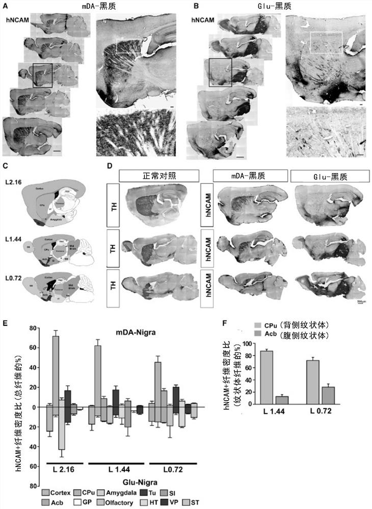
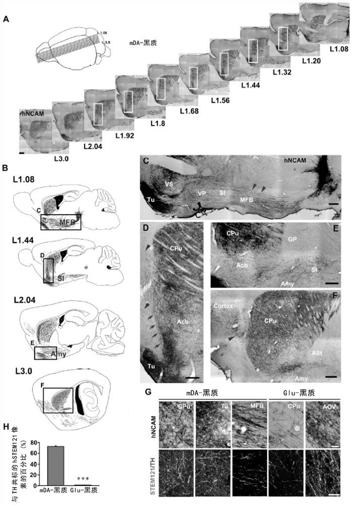
[0235] From serial sagittal sections ( figure 2 A) shows that transplanted mDA neurons extend rostral axons along the well-defined medial forebrain bundle (MFB) ( figure 2 B, L1.08 and the red arrow at figure 2 C). Viewed from the side, they extend across SI ( figure 2 B, L1.44) and amygdala ( figure 2 B, L2.04). Viewed from the rostral side of the beak, hNCAM+ fibers passed through the Acb( figure 2 B-2E), where most of the hNCAM+ fibers enter the CPu along the border between cortex and striatum ( figure 2 A and figure 2 red arrow in D). Few hNCAM+ fibers were detected in the cortex ( figure 2 Blue arrows in D and 2F). These results indicate that most of the axonal projections of transplanted mDA neurons follow the endogenous substantia nigra-striatum pathway.
[0236] In the dorsal / lateral striatum, the dense hNCAM+ fibers were branched, forming a dense branched network...
Embodiment 3
[0238] Example 3. Genetic markers reveal specific axonal innervation of human mDA neurons
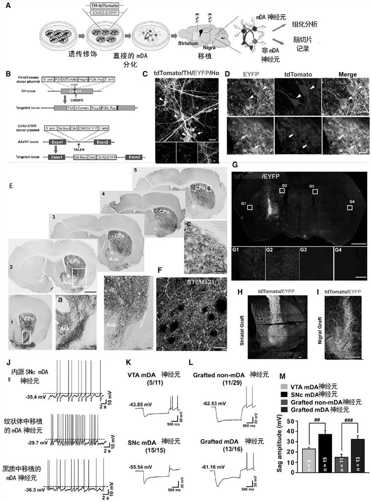
[0239] To elucidate the specific axonal innervation elicited by transplanted mDA neurons, the inventors established a TH reporter hESC line with tdTomato expression recapitulating the expression of the endogenous TH gene (Methods Details). The inventors further knocked the ChR2-EYFP fusion protein expression cassette into the AAVS1 gene locus to realize specific markers and manipulation of transplanted human cells ( image 3 A, 3B, 10A and 10B). The final hESC, termed TH-tdTomato / AAVS1-ChR2-EYFPhESC, constitutively expressed ChR2-EYFP throughout mDA neuronal differentiation, whereas tdTomato was expressed only at later stages of mDA neuronal differentiation ( Figure 10 C), and is expressed only in TH+ mDA neurons (Figure 3C and 10D), emphasizing TH+ cell-specific expression.
[0240] Then, the inventors transplanted mDA precursor cells derived from TH-tdTomato / AAVS1-ChR2-EYFP hESCs i...
PUM
 Abstract
Description
Claims
Application Information
 - R&D Engineer
- R&D Manager
- IP Professional
- Industry Leading Data Capabilities
- Powerful AI technology
- Patent DNA Extraction
Browse by: Latest US Patents, China's latest patents, Technical Efficacy Thesaurus, Application Domain, Technology Topic, Popular Technical Reports.
© 2024 PatSnap. All rights reserved.Legal|Privacy policy|Modern Slavery Act Transparency Statement|Sitemap|About US| Contact US: help@patsnap.com