
Sealing cap used for endoscope and ureter or flow guide tube in matched mode

A technology of sealing cap and ureter, which is applied in the field of medical devices, can solve problems such as inconvenient position adjustment of endoscopes, and achieve the effects of improved convenience, low manufacturing cost, and simple structure

Active Publication Date: 2022-04-12
姜玉雷
View PDF6 Cites 0 Cited by
  • Summary
  • Abstract
  • Description
  • Claims
  • Application Information

AI Technical Summary

Problems solved by technology

[0005] The technical problem to be solved by the present invention is: to solve the disadvantages that it is inconvenient to adjust the position of the cavity mirror after it is fixed, and it is impossible to fix or adjust the cavity lens in one direction

Method used


Image

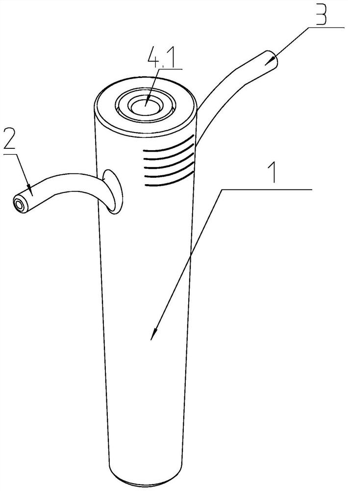
  • Sealing cap used for endoscope and ureter or flow guide tube in matched mode
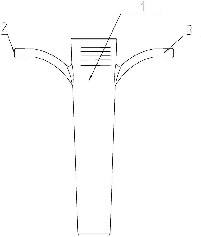
  • Sealing cap used for endoscope and ureter or flow guide tube in matched mode
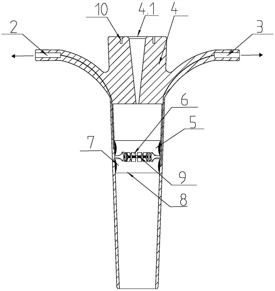
  • Sealing cap used for endoscope and ureter or flow guide tube in matched mode

Examples


Embodiment approach 1

[0050] (refer to Figure 6-Figure 7 ) inflate the first inflatable part 5 through the first inflatable channel 2, and inflate the second inflatable part 7 through the second inflatable channel 3, so that the first inflatable part 5 is inflated to form a first balloon, and the second inflatable part is inflated Part 7 forms a second balloon, the first balloon and the second balloon expand at the same time, and the expansion of the walls of the two balloons generates outward tension, leaning against both sides of the limiting part 6, and the limiting part is supported by the two balloons. 6 has a certain bending strength, so that the limiting part 6 cannot swing upwards or downwards. It is equivalent to that the clamping limit 6 is clamped on the outer wall of the cavity mirror 11 with a certain strength. Furthermore, in the process, when the endoscope 11 needs to be fixed, the stopper 6 can apply a radial force around the tube wall of the endoscope 11 to increase the frictiona...

Embodiment approach 2

[0052] (refer to Figure 8-Figure 9 ) only inflate the first inflatable part 5 through the first inflatable channel 2, so that the first inflatable part 5 expands to form the first balloon, and the first balloon wall expands to generate outward tension, so that the first balloon leans against the limit At this time, the limiting portion 6 can only bend downward. If the limiting part 6 bends downward, the diameter of the space enclosed by the end of the limiting part 6 will decrease, which is beneficial for the endoscope 11 to pass through when inserted, that is, the endoscope 11 can only move in the "extending" direction. However, since the first balloon leans against the limiting portion 6, when the endoscope 11 has passed the limiting portion 6 and is pulled out, the limiting portion 6 cannot bend upwards, and the endoscope 11 will be clamped on the limiting portion 6, therefore, the movement of the cavity mirror 11 in the "retracting" direction is limited. This can facili...

Embodiment approach 3

[0054] (refer to Figure 10-Figure 11 ) This embodiment is opposite to Embodiment 2. The second inflatable part 7 is only inflated to form the second balloon, so that the second balloon leans against the bottom of the limiting part 6. At this time, the limiting part 6 can only bend upwards, and the endoscope 11 can only carry out the movement of " shrinking " direction. Therefore, the movement of the endoscope 11 in the "extend" direction is limited. This can facilitate the diagnosis and treatment of the endoscope 11 only to the outside, and is generally used for ureteral calculus excretion.


PUM


Abstract

The invention provides a sealing cap for cooperative use of an endoscope and a ureter or a flow guide tube, which comprises a first inflation channel arranged on a sealing cap body and used for inflating a first inflation part, and a first balloon capable of extruding a limiting part to one side to bend the limiting part, and a second inflation channel arranged on the sealing cap body and used for inflating the first inflation part to form a second balloon capable of extruding the limiting part to one side to bend the limiting part. And the second inflation channel is used for inflating the second inflation part, so that the second inflation part can form a second balloon which extrudes the pressure limiting part towards the other side to be bent. The device has the beneficial effects that the air bags capable of being contracted and expanded are arranged on the two sides of the limiting part correspondingly, the setting angle of the end of the limiting part can be restrained through expansion or contraction of the different air bags, and the limiting part is promoted to swing up and down according to the use requirement; therefore, the full fixation of the telescopic freedom degree of the endoscope and the unidirectional freedom degree fixation are realized.

Description

technical field [0001] The invention relates to the field of medical instruments, in particular to a sealing cap used in conjunction with a laparoscope and a ureter or a drainage tube. Background technique [0002] Laparoscopy can clearly observe the lesions in the kidney or ureter, such as stones, tumors, etc., to diagnose and treat diseases in the cavity. The existing laparoscopy requires a catheter or drainage tube to enter the renal calices to observe the kidney. Diagnosis and treatment of diseases of the upper ureter. The matching position between the endoscope and the urinary catheter or drainage tube needs to be sealed and connected with a waterproof sealing cap. The waterproof sealing cap prevents the liquid and gas in the cavity from flowing out from the gap between the endoscope and the urinary catheter or drainage tube. [0003] In the current operation, the endoscope is usually fixed at a specific position by hand for observation and treatment. Holding the endos...

Claims


Application Information

Patent Timeline
Patent Type & Authority Applications(China)
IPC IPC(8): A61M39/20
Inventor 姜玉雷
Owner 姜玉雷
Learn More