
System for realizing oral cavity shaping simulation display by combining 3D image with feature fusion technology

A feature fusion and display system technology, applied in 3D modeling, image analysis, image enhancement, etc., can solve problems such as viewing dead angles, difficult to see the oral cavity, and increasing the difficulty for doctors to view oral diseases

Active Publication Date: 2022-05-10
西安交通大学口腔医院
View PDF10 Cites 0 Cited by
  • Summary
  • Abstract
  • Description
  • Claims
  • Application Information

AI Technical Summary

Problems solved by technology

[0003] At present, when viewing the oral cavity, most doctors directly observe the patient's oral cavity, and there are dead spots in the viewing, and it is difficult to observe the internal conditions of the oral cavity, and In the later study of the patient's disease, it is also impossible to make a sound description. At the same time, when the patient has tone, it will increase the difficulty for the doctor to watch the patient's oral disease.

Method used


Image

  • System for realizing oral cavity shaping simulation display by combining 3D image with feature fusion technology
  • System for realizing oral cavity shaping simulation display by combining 3D image with feature fusion technology
  • System for realizing oral cavity shaping simulation display by combining 3D image with feature fusion technology

Examples


Embodiment 1

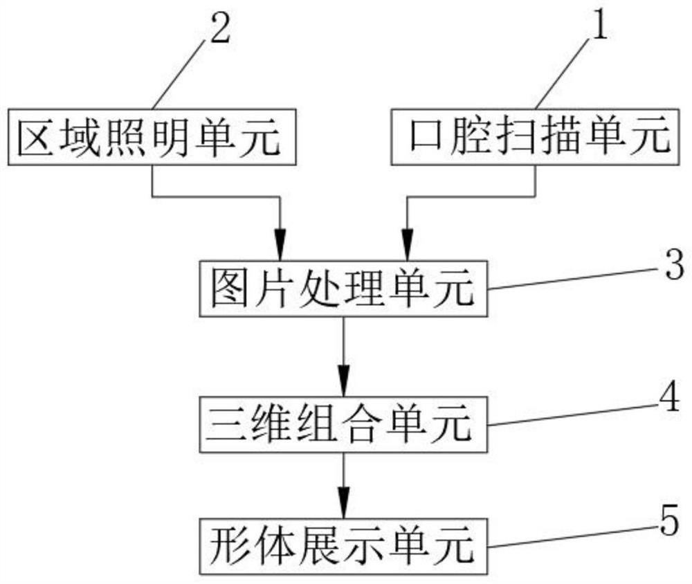
[0066] The present invention provides a 3D image combined with feature fusion technology to realize oral cavity shaping simulation display system, please refer to Figure 1-Figure 7 , including an oral cavity scanning unit 1, an area lighting unit 2, an image processing unit 3, a three-dimensional combination unit 4 and a body display unit 5;

[0067] The oral cavity scanning unit 1 is used to scan the inside of the patient's oral cavity to capture the internal situation of the patient's oral cavity, so that the conditions of the teeth and oral lining in the patient's oral cavity are displayed for others to watch;

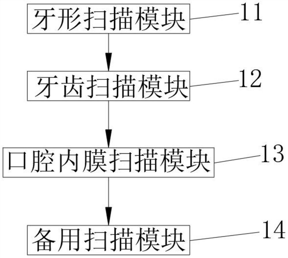
[0068] The oral cavity scanning unit 1 includes a tooth shape scanning module 11, a tooth scanning module 12 and an oral cavity endometrial scanning module 13;

[0069] Tooth shape scanning module 11 is used for scanning and photographing the shape of the teeth in the patient's oral cavity to obtain the shape of the patient's teeth, and when modeling the patient's ...

Embodiment 2

[0118] Considering that in the process of screening the pictures in the later stage, there is a place where the image is blank due to the unclear shooting, so it cannot be modeled normally, refer to figure 2 , the oral cavity scanning unit 1 also includes a backup scanning module 14, the backup scanning module 14 is used to scan and capture a place in the patient's oral cavity multiple times;

[0119] The spare scanning module 14 scans and captures a certain part of the oral cavity multiple times to ensure that the features are not clearly displayed in the process of later image processing, so as to ensure the accuracy of image shooting.


PUM


Abstract

The invention relates to the technical field of 3D image shaping simulation, in particular to a 3D image and feature fusion technology combined oral cavity shaping simulation display system. The system comprises an oral cavity scanning unit, an area lighting unit, a picture processing unit, a three-dimensional combination unit and a shape display unit. The oral cavity scanning unit scans and captures the shape of the oral cavity of a patient and the internal condition of the oral cavity, and the picture processing unit and the three-dimensional combination unit process and model the picture and the oral cavity structure, so that the oral cavity condition of the patient is displayed in front of the eyes of a doctor; the problem of watching dead angles caused by watching the oral cavity of the patient is avoided, so that a doctor can clearly know the oral cavity problem of the patient, and the doctor can conveniently take medicine according to the situation of the patient.

Description

technical field [0001] The invention relates to the field of 3D image shaping simulation technology, in particular to a 3D image combined with feature fusion technology to realize oral cavity shaping simulation display system. Background technique [0002] 3D images have the advantages of three-dimensionality and easy viewing. They are often used in parts design, product development, and medicine. In medicine, 3D images are often used to describe patients’ illnesses and speculate on patients’ treatment methods for convenience. The understanding of other doctors and the application of 3D images in medicine are also convenient for doctors to teach other doctors and enhance the learning effect of doctors. [0003] At present, when viewing the oral cavity, most doctors directly observe the patient's oral cavity, and there are blind spots in the viewing. It is difficult to observe the internal conditions of the oral cavity, and it is impossible to make a sound description in the ...

Claims


Application Information

Patent Timeline
IPC IPC(8): G06T7/593G06T7/136G06V10/44G06T17/00
CPCG06T7/593G06T7/136G06T17/00G06T2207/10012G06T2207/30036
Inventor 刘飞
Owner 西安交通大学口腔医院
Learn More