
Cardiovascular ultrasonic diagnosis device

A technology of ultrasonic diagnosis and diagnostic device, which is applied in the direction of ultrasonic diagnosis, infrasonic diagnosis, ultrasonic/sonic/infrasonic diagnosis, etc. It can solve problems such as loose parts, poor dustproof and anti-collision effects, and damage to ultrasonic diagnostic devices, so as to reduce the Occupies space, improves fixation effect, and improves practicality

Inactive Publication Date: 2022-07-12
临颍县人民医院
View PDF0 Cites 0 Cited by
  • Summary
  • Abstract
  • Description
  • Claims
  • Application Information

AI Technical Summary

Problems solved by technology

[0004] The prior art has the following disadvantages: when the ultrasonic diagnostic device in the prior art is transported or moved, due to the poor shock absorption effect, the internal parts of the ultrasonic diagnostic device are often loosened or damaged due to large vibrations. Most of them are placed outside in the open air, and the dustproof and anti-collision effects are poor. When there is a lot of dust entering the ultrasonic diagnostic device, the service life of the ultrasonic diagnostic device will be greatly reduced, and the ultrasonic diagnostic device is easily damaged when it is impacted.

Method used


Image

  • Cardiovascular ultrasonic diagnosis device
  • Cardiovascular ultrasonic diagnosis device
  • Cardiovascular ultrasonic diagnosis device

Examples


Embodiment Construction

[0026] The present invention will be further described below with reference to the accompanying drawings and examples, and the modes of the present invention include but are not limited to the following examples.

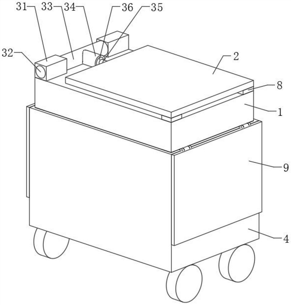
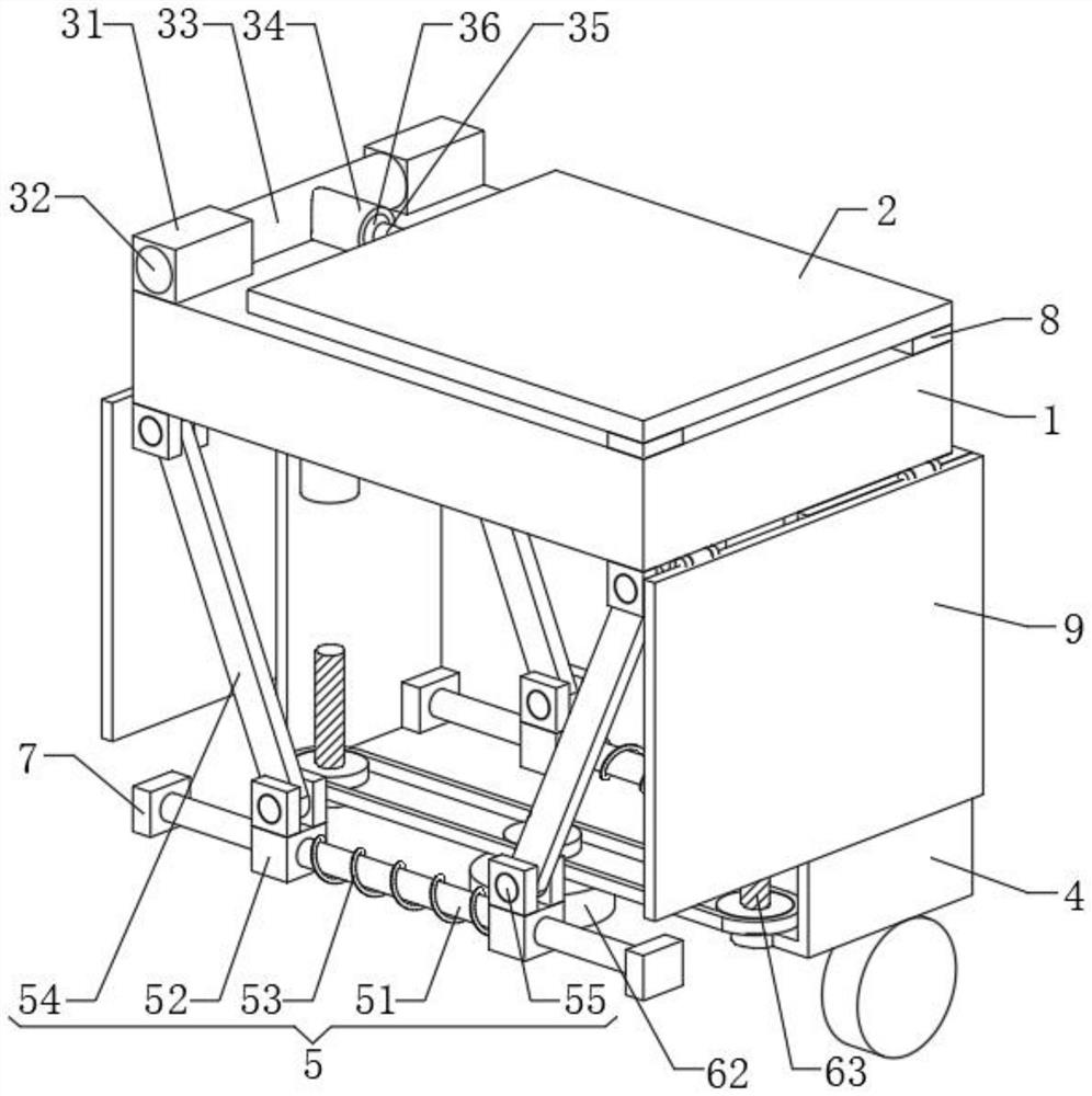
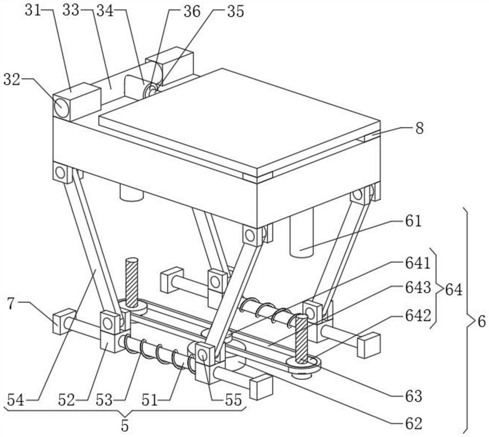
[0027] Please refer to the attached manual Figure 1 to Figure 7 As shown, a cardiovascular ultrasonic diagnostic apparatus includes an ultrasonic diagnostic apparatus body 1 and a display screen 2 arranged on the top of the ultrasonic diagnostic apparatus main body 1. The bottom of the ultrasonic diagnostic apparatus body 1 is provided with a cavity 4, and the inner cavity of the cavity 4 has two inner cavities. Both ends are provided with an elastic mechanism 5, and a fixing mechanism 6 is provided between the ultrasonic diagnostic device body 1 and the cavity 4; It is fixed on the top of the cavity 4 with the elastic mechanism 5;

[0028] By adding an elastic mechanism 5 in the cavity 4, the ultrasonic diagnostic device body 1 can be buffered, and when the ultra...


PUM


Abstract

The invention discloses a cardiovascular ultrasonic diagnosis device which comprises an ultrasonic diagnosis device body and a display screen arranged at the top of the ultrasonic diagnosis device body, and a cavity is formed in the bottom of the ultrasonic diagnosis device body. According to the ultrasonic diagnosis device, the elastic mechanism is additionally arranged in the inner cavity of the cavity, the ultrasonic diagnosis device body can be buffered, when the ultrasonic diagnosis device body is transported or moved, the ultrasonic diagnosis device body is effectively prevented from being damaged due to large vibration, and when the ultrasonic diagnosis device body is not used, the ultrasonic diagnosis device body is prevented from being damaged. The ultrasonic diagnosis device body is fixed to the top of the inner cavity of the cavity through the fixing mechanism, the ultrasonic diagnosis device body is stored through the cavity, the fixing stability of the fixing mechanism to the ultrasonic diagnosis device body can be improved through the elastic mechanism, and the ultrasonic diagnosis device body can be subjected to dustproof treatment and protection treatment; and the occupied space of the ultrasonic diagnosis device body can be effectively reduced, the inclination angle and the orientation of the display screen are adjusted through the adjusting mechanism, and therefore the practicability of the display screen can be improved.

Description

technical field [0001] The invention relates to the field of ultrasonic diagnostic devices, in particular to a cardiovascular ultrasonic diagnostic device. Background technique [0002] The heart is a hollow muscular organ located in the middle of the thoracic cavity and divided into left and right chambers by a septum, and each chamber is further divided into two parts, the atrium in the upper part and the ventricle in the lower part. The atria collect blood into the heart, and the ventricles eject blood out of the heart. The ventricles have valves at the inlet and outlet, which keep blood flowing in one direction. Under different physiological conditions of the human body, the metabolic levels of various organs and tissues are different, and the needs for blood flow are also different. Cardiovascular activity can change cardiac output and peripheral resistance under the mediation of the body's nerves and body fluids, and coordinate the distribution of blood flow among va...

Claims


Application Information

Patent Timeline
Patent Type & Authority Applications(China)
IPC IPC(8): A61B8/08A61B8/00F16M11/04F16M11/14F16M11/18F16M11/42
CPCA61B8/0883A61B8/0891A61B8/44A61B8/462F16M11/04F16M11/14F16M11/18F16M11/42
Inventor 彭新沛陈鹏丽
Owner 临颍县人民医院
Learn More