X-ray computed tomographic apparatus, image processing apparatus, and image processing method

An image processing device and image processing technology, applied in image data processing, image data processing, calculation and other directions, can solve the problem that the location and location of coronary artery lesions cannot be determined.

Active Publication Date: 2004-11-03
TOSHIBA MEDICAL SYST CORP
View PDF0 Cites 13 Cited by
  • Summary
  • Abstract
  • Description
  • Claims
  • Application Information

AI Technical Summary

Problems solved by technology

[0005] By displaying the pole map superimposed on the typical dominant section as described above, it can be understood which of the left anterior descending artery, the left circumflex artery, and the right coronary

Method used


Image

  • X-ray computed tomographic apparatus, image processing apparatus, and image processing method
  • X-ray computed tomographic apparatus, image processing apparatus, and image processing method
  • X-ray computed tomographic apparatus, image processing apparatus, and image processing method

Examples


Embodiment Construction

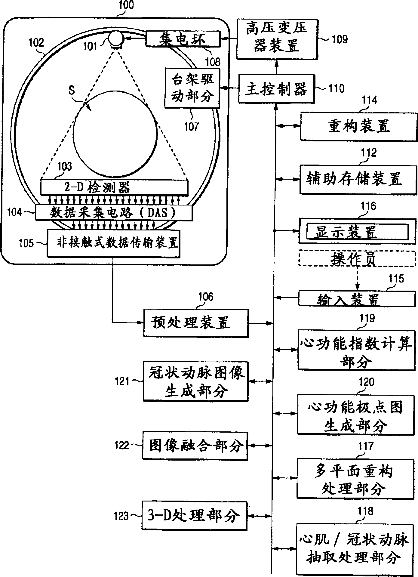
[0030] The X-ray computed tomography apparatus (X-ray CT scanner), image processing apparatus, and image processing method of the present invention will be described below with reference to the drawings. In this embodiment, an X-ray CT scanner will be described. However, the X-ray CT scanner described below is equipped with an image processing device having a function of realizing the image processing method of the present embodiment.

[0031] By the way, X-ray CT scanners include various types such as rotary / rotary type, in which the X-ray tube and radiation detector are integrally rotated around the subject; fixed / rotary type, in which a plurality of detection units are arranged in a circular array, and The X-ray tube alone rotates around the subject; and the invention is applicable to either type. Furthermore, to reconstruct the tomographic data of a slice requires projection data of the entire circle, ie about 360° around the object, whereas projection data of 180° plus v...


PUM


Abstract

An image processing apparatus includes an index computing unit that computes motion functional indices of a particular organ from volume data of a subject including the particular organ, a polar map generating unit that generates a polar map by developing the indices on a polar coordinate system, an image generating unit that generates, from the volume data, an image related to a structure of one of the particular organ and a peripheral organ developed on the polar coordinate system, a fusing unit that fuses the image into the polar map, and a display unit that displays a polar map fused with the image.

Description

technical field [0001] The present invention relates to an X-ray computed tomographic apparatus, an image processing apparatus and an image processing method for generating a pole map related to a functional index of an organ such as the heart. Background technique [0002] The accelerated development of helical scanning and the development of volume scanning using two-dimensional detectors have greatly changed the concept of X-ray computed tomography devices from simple cross-sectional imaging devices to volume imaging devices. In other words, X-ray computed tomography apparatus employing these scanning methods can perform fast volume scanning of objects with a short temporal resolution. The X-ray computed tomography apparatus equipped with this function finds practical use in many diagnostic fields where the X-ray computed tomography apparatus has not been used originally and efforts are being made to find new diagnostic methods. [0003] One of the possible directions is...

Claims


Application Information

Patent Timeline
IPC IPC(8): A61B6/03G06T1/00G06T3/00
CPCA61B6/5235A61B6/466A61B6/032A61B6/504A61B6/463A61B6/503
Inventor 藤泽恭子
Owner TOSHIBA MEDICAL SYST CORP
Why Eureka
Try Eureka