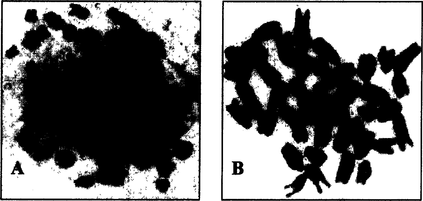
Sub-clone cell line mestastatic-human liver cancer and establishing method
A technology for liver cancer metastasis and liver cancer cells, applied in the field of animal cell lines, can solve the problems of lack of humoral and cellular immune functions, loss of T and B lymphocyte immune functions, etc.
- Summary
- Abstract
- Description
- Claims
- Application Information
AI Technical Summary
Problems solved by technology
Method used
Image
Examples
Embodiment 1
[0035] A method for establishing a human liver cancer metastasis cell line, comprising the steps of:
[0036] (1) Inoculation of SCID mice: Get 5 SCID mice, male or female, with body weights of 13.1g, 14.3g, 14.4g, 15.2g, and 15.8g respectively, aged 4-7 weeks, and raised in a special pathogen-free environment. The human liver cancer cell H7402 in the logarithmic growth phase was made into a 0.2ml single-cell suspension containing 1×10 7 cells, seeded on Cs 137 γ-ray irradiated SCID rats subcutaneous in the axillae and back, SCID rats were provided by Tianjin Institute of Hematology, Chinese Academy of Medical Sciences;
[0037] (2) Confirmation of the metastatic site: the tumor-bearing SCID mice were killed by necking, and the pathological sections of each tissue were observed under a microscope to determine that the metastatic site was the lung;
[0038] (3) Primary culture: On the 66th day after inoculation of human hepatoma cell H7402, the tumor-bearing SCID mice were ki...
Embodiment 2
[0063] A method for establishing a human liver cancer metastasis cell line, comprising the steps of:
[0064] (1) Inoculation of SCID mice: take 4 SCID mice, male or female, weighing 13.4g, 13.6g, 14.3g, 15.8g respectively, aged 4-7 weeks, raised in a special pathogen-free environment, and will grow logarithmically 0.2ml single-cell suspension containing 1×10 7 cells, seeded on Cs 137 γ-ray irradiated SCID rats subcutaneous in the axillae and back, SCID rats were provided by Tianjin Institute of Hematology, Chinese Academy of Medical Sciences;
[0065] (2) Confirmation of the metastatic site: the tumor-bearing SCID mice were killed by necking, and the pathological sections of each tissue were observed under a microscope to determine that the metastatic site was the lung;
[0066] (3) Primary culture: On the 55th day after inoculation of human hepatoma cell H7402, tumor-bearing SCID mice were killed by necking, aseptically dissected, and the lungs were taken out for primary c...
Embodiment 3
[0070] A method for establishing a human liver cancer metastasis cell line, comprising the steps of:
[0071] (1) Inoculation of SCID mice: Take 3 SCID mice, male or female, weighing 14.4g, 15.3g, 15.7g, aged 4-7 weeks, raised in a special pathogen-free environment, and inoculate human liver cancer cells in logarithmic growth phase H7402 made into 0.2ml single cell suspension containing 1×10 7 cells, seeded on Cs 137 γ-ray irradiated SCID rats subcutaneous in the axillae and back, SCID rats were provided by Tianjin Institute of Hematology, Chinese Academy of Medical Sciences;
[0072] (2) Confirmation of the metastatic site: the tumor-bearing SCID mice were killed by necking, and the pathological sections of each tissue were observed under a microscope to determine that the metastatic site was the lung;
[0073](3) Primary culture: On the 67th day after inoculation of human hepatoma cell H7402, the tumor-bearing SCID mice were killed by necking, aseptically dissected, and th...
PUM
 Abstract
Description
Claims
Application Information
 - R&D
- Intellectual Property
- Life Sciences
- Materials
- Tech Scout
- Unparalleled Data Quality
- Higher Quality Content
- 60% Fewer Hallucinations
Browse by: Latest US Patents, China's latest patents, Technical Efficacy Thesaurus, Application Domain, Technology Topic, Popular Technical Reports.
© 2025 PatSnap. All rights reserved.Legal|Privacy policy|Modern Slavery Act Transparency Statement|Sitemap|About US| Contact US: help@patsnap.com


