Holographic cardiovector three-dimensional image display instrument

An ECG vector and three-dimensional image technology, applied in the field of analysis and diagnosis systems, can solve problems such as low intensity, lack of ECG signals, and no three-dimensional image display, and achieve the effect of improving reliability

Inactive Publication Date: 2005-07-20
徐文廷
View PDF0 Cites 5 Cited by
  • Summary
  • Abstract
  • Description
  • Claims
  • Application Information

AI Technical Summary

Problems solved by technology

This lack of ECG signals in the upper and lower intervals has obvious limitations in the amount of information
[0006] The second is the lack of intuitive three-dimensionality
The more serious so-called simulated ECG three-dimensional graphics are more indirect, more virtual, and have greater errors
[0007] The third is that it does not show its essence in the form of stereoscopic images that it originally had.
CT and B-ultrasound imaging are the attenuation or reflection changes of external signals in the human body, and are obtained through data processing, but ECG signals are generated by the human body itself, and the intensity is not low, so it should be easier to display in a three-dimensional form, but now There are instruments that can't do it

Method used


Image

  • Holographic cardiovector three-dimensional image display instrument
  • Holographic cardiovector three-dimensional image display instrument
  • Holographic cardiovector three-dimensional image display instrument

Examples


Embodiment Construction

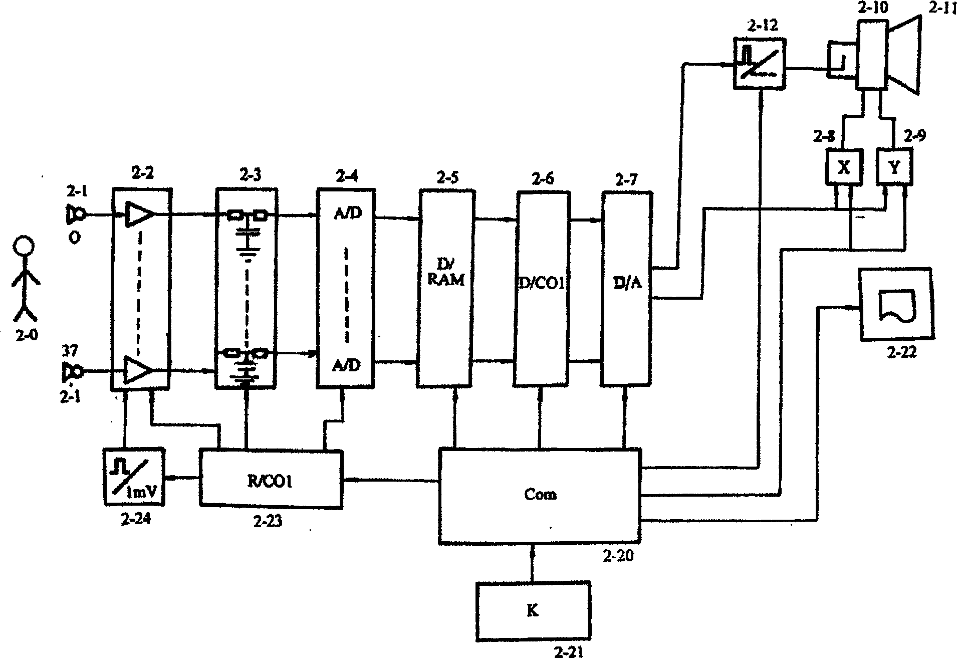
[0017] figure 2 Shown is a fully self-contained embodiment. The detection electrode 2-1, the ECG signal detected from the subject is pre-amplified by the preamplifier 2-2, filtered by the band-pass filter 2-3 to eliminate interference, and passed by the analog / digital signal converter 2-4 , converting the analog ECG signal into a digital signal, and then sending it into the digital memory 2-5 for storage. Under the control of the computer 2-20, the data of different leads are sequentially selected through the data strobe 2-6, and the digital quantity of the signal is converted into an analog quantity through the digital / analog signal converter 2-7, and then the digital quantity of the signal is converted into an analog quantity through the display screen. The x, y deflection drive systems 2-8, 2-9 drive the deflection control part 2-10 of the picture tube 2-11, so as to realize the graphics of the electrocardiogram vector bar or vector tube displayed on the screen 2-11.

[...


PUM


Abstract

The present invention relates to a homographic vectorcardiographic three-dimensional image displaying instrument. It has three structure modes: first mode mainly is formed from detection electrode, preamplifier, A/D converter, memory, D/A converter, display, computer and printer, its dat a processing is simplemented by software operation; its second mode is formed from a cardioelectric functional component cardioelectric working station and comptuer and correspondent software; and its third mode is composed of several cardioelectric functional component cardioelectric working stations and computer and correspondent software. Said invention also provides its working principle.

Description

technical field [0001] The invention is an electrocardiographic analysis and diagnosis device, and particularly belongs to the technical field of an analysis and diagnosis system capable of displaying electrocardiographic vectors in a holographic three-dimensional image manner. Background technique [0002] All kinds of electrocardiographic examination and diagnostic instruments are actually a kind of measurement and verification device, no matter whether these instruments include intelligent components or not, and how complete and accurate the performance of the instrument itself is, but in terms of clinical application, its The accuracy or reliability of the detection is the worst compared with the same quality instruments in other fields. The reason is not that the accuracy of the instrument itself is not enough, but that there are deficiencies in the detection method, so that in serious cases, the electrocardiogram of a patient with severe heart disease a few minutes bef...

Claims


Application Information

Patent Timeline
Patent Type & Authority Applications(China)
IPC IPC(8): A61B5/0402A61B5/044
Inventor 徐文廷徐凌宇徐怀宇徐淑英唐文晶郭非凡苏坤祥
Owner 徐文廷
Why Eureka
Try Eureka