
Method of producing tomosynthesis image of beating heart and tomosynthesis device

A technology of tomography and heart, applied in computerized tomography scanner, diagnosis, echo tomography, etc., can solve the problems of high radiation burden of patients, EKG has no shape, etc.

Inactive Publication Date: 2006-08-16
SIEMENS AG
View PDF0 Cites 1 Cited by
  • Summary
  • Abstract
  • Description
  • Claims
  • Application Information

AI Technical Summary

Problems solved by technology

[0003] Problems can arise, for example, when using the EKG signal to determine the resting phase of the heart that the described EKG does not have a typical shape due to stimulation pathway disturbances or prominent infarcts, making automatic or manual selection of the correct phase segment problematic
[0004] Although some methods were also proposed during this period in which the current state of motion of the heart can be deduced from the detector information, they required the patient to be scanned over the entire cardiac cycle, resulting in a high patient sensitivity, at least in computed tomography examinations. Radiation burden

Method used


Image

  • Method of producing tomosynthesis image of beating heart and tomosynthesis device

Examples


Embodiment Construction

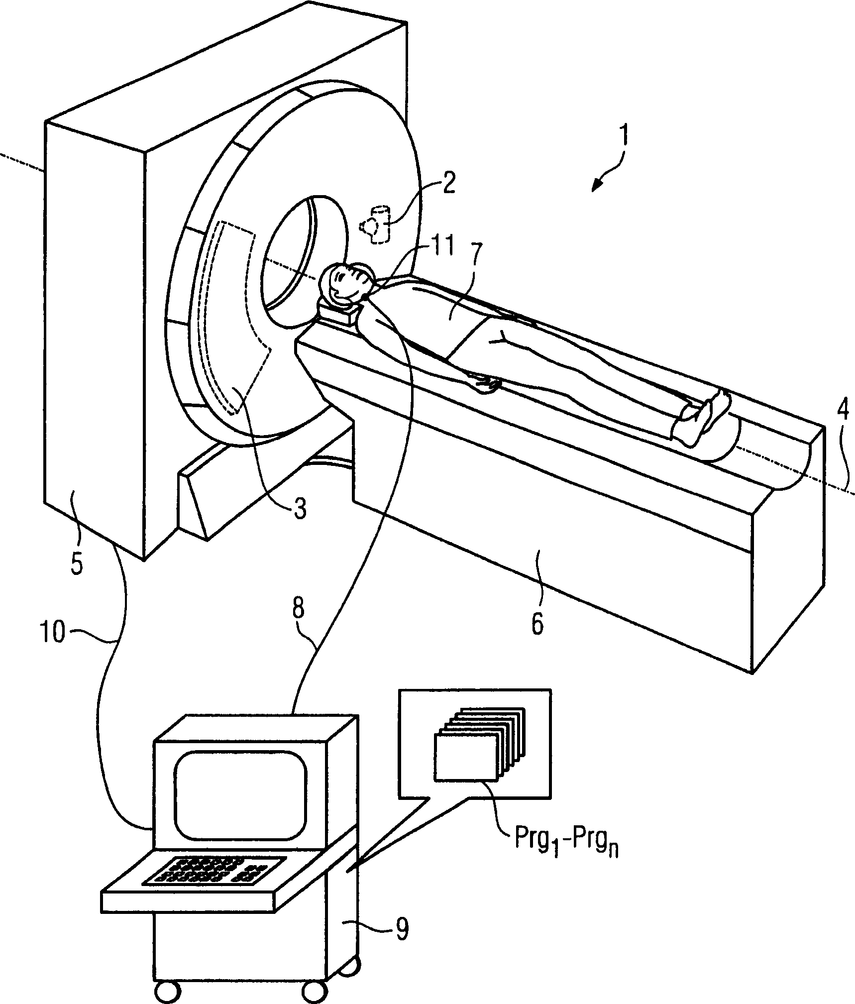
[0023] figure 1 A computed tomography system 1 is shown with a housing 5 , in which a support with a circularly running x-ray tube 2 and a plurality of rows of detectors 3 arranged opposite it can be seen. Furthermore, a patient 7 is shown on a patient couch 6 , which can be moved into the CT1 opening during the scanning process. During the scanning process, the X-ray tube 2 moves circularly around the patient 7, which can make the patient 7 move relatively along the direction of the system axis 4, thereby performing a helical scan relative to the patient 7, or by making the patient 7 move in the direction of the X-ray tube The patient is scanned by sequentially moving forward during scan intervals in a plurality of pure circular motions relative to the patient. The computer tomography system 1 is controlled by a control and computing unit 9 via a control / data line 10 . The data collected by the detector 3 is also transmitted to the computer via the control / data line 10 . T...


PUM


Abstract

A method and a tomography unit are disclosed for taking tomographic pictures of a patient's beating heart, in particular X-ray CT pictures. In such a method and tomography unit, only detector data or image data of a portion of the cardiac phase from one or more heart beats is used for taking a picture. Further, at least one pressure signal, produced by the mechanical cardiac pulse and varying temporally with the cardiac cycle, is used for determining the cardiac phase.

Description

technical field [0001] The invention relates to a method and a tomography system for generating a tomographic image, in particular an X-ray CT image, of a beating patient's heart, in which only the detection of partial cardiac phases in one or more heart beats is used to generate the image device data or image data. Background technique [0002] Similar methods are generally known. For example when generating computed tomography images, record the EKG signal while scanning the beating heart and select the resting phase of the heart based on the EKG signal, especially the R tooth in the EKG signal, and use the detector data only for this cycle phase or image data and integrate multiple heart beats, thereby reconstructing or combining high-quality images. [0003] Problems can arise, for example, when using the EKG signal to determine the resting phase of the heart that the described EKG does not have a typical shape due to stimulation pathway disturbances or prominent infar...

Claims


Application Information

Patent Timeline
IPC IPC(8): A61B6/03
CPCA61B6/503A61B6/541
Inventor 赫伯特·布鲁德
Owner SIEMENS AG
Learn More